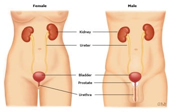
Another important function of the kidneys is electrolyte handling, specifically phosphorus and potassium. When the kidneys are not functioning well, both potassium and phosphorus can build up in the blood. When the phosphorus is elevated, the calcium in your bones is decreased resulting in weak bone. Elevated potassium in the blood can cause problems with your heart rhythm and at certain levels is a medical emergency requiring ER evaluation.
The toxins or waste in the blood comes from the normal breakdown of muscle and the food you eat. Food is used by your body for self-repair and energy. Afterward, the waste is moved to the blood. When the kidneys are not working to remove the wastes, they would build up in the blood and can cause damage.
The filtering occurs inside your kidney. There are tiny units called nephrons. Each kidney has about 1 million nephrons. Here small blood vessels called capillaries combine with urine-carrying tubes known as tubules. Then a complicated chemical exchange happens, waste materials and excess water leave the blood and enter into the urinary system. The kidney then measures out electrolytes, such as phosphorus, sodium, and potassium and then releases them back to the blood.
There are several important hormones released from the kidney. Erythropoietin stimulates red blood cell production in the bones. Renin, regulates blood pressure and the active form of vitamin D helps maintain calcium for the bones and normal balance in the body.
The most common causes of kidney disease are hypertension and diabetes. Diabetes prevents your body from using sugar appropriately. When sugar stays in the blood instead of breaking down, it can become a poison. This causes damage to the kidney and is called diabetic nephropathy.
Uncontrolled blood pressure may cause damage to the small blood vessels in the kidney. When this happens, the damaged vessels are unable to filter poison from the blood. Your nephrologist may prescribe medications to treat the hypertension. There is a group of medications used to treat hypertension called ACE inhibitors that may give extra protection in patients with diabetes.
Some kidney diseases are inherited. Polycystic kidney disease, for example, is a genetic disorder that causes cysts grow in the kidneys. These cysts can slowly replace much of the mass of the kidneys, causing reduced kidney function and leading to kidney failure.
Some other causes are due to poisons or trauma, A direct forceful blow to the kidneys, may lead to kidney disease. Many over-the-counter medications may harm your kidneys when taken regularly over a long time. These medications often combine aspirin, acetaminophen, and other medications like ibuprofen can be dangerous to the kidney.
Unfortunately, there is not a cure for renal failure. However, if you are in the early stages of the disease, you may be able to slow the progression of disease and make the kidneys last longer.
– If you have diabetes, watch your blood sugar closely to keep it under control.
-Have your blood pressure checked regularly. Eat healthy diet with low salt
-Avoid pain pills that may make your kidney disease worse.
- CKD Stage 1 — GFR > 90
- CKD Stage 2 — GFR 60-89
- CKD Stage 3 — GFR 30-59
- CKD Stage 4 — GFR 15-29
- CKD Stage 5 — GFR < 15 or Dialysis
Urine Tests – Urinalysis, protein/creatinine ratio
Imaging Tests – A renal ultrasound may be completed to assess the size, shape and anatomy of your kidney.
Kidney Biopsy – A kidney biopsy is a test where a small piece of kidney tissue is removed by a needle. The tissue is examined under a microscope to determine the cause of kidney disease.
Hemodialysis – This modality cleans your blood using a machine with a filter called a dialyzer. During a hemodialysis treatment blood travels from your body through tubes to the dialyzer which filters out wastes and extra water. The cleaned blood flows through another set of tubes back into your body. This is usually performed in a dialysis unit three times a week for 4 hours each treatment.
Peritoneal Dialysis – removes wastes and extra water from your body using the lining of your abdomen to filter your blood. A solution travels through a soft tube into your abdomen. The solution draws wastes and extra water from tiny blood vessels in your abdomen back into the solution which is then drained from your abdomen through the soft tube. This form of dialysis is completed at your home every night.
| Zip Code | City | County |
| 36201 | Anniston | Calhoun |
| 36202 | Anniston | Calhoun |
| 36203 | Anniston | Calhoun |
| 36203 | Oxford | Calhoun |
| 36204 | Anniston | Calhoun |
| 36204 | Blue Mountain | Calhoun |
| 36205 | Fort McClellan | Calhoun |
| 36205 | Anniston | Calhoun |
| 36206 | Anniston | Calhoun |
| 36207 | Anniston | Calhoun |
| 36250 | Alexandria | Calhoun |
| 36253 | Bynum | Calhoun |
| 36254 | Choccolocco | Calhoun |
| 36257 | De Armanville | Calhoun |
| 36260 | Eastaboga | Calhoun |
| 36265 | Jacksonville | Calhoun |
| 36271 | Ohatchee | Calhoun |
| 36272 | Borden Springs | Calhoun |
| 36272 | Piedmont | Calhoun |
| 36277 | Weaver | Calhoun |
| 36279 | Wellington | Calhoun |
| 35013 | Allgood | Blount |
| 35031 | Blountsville | Blount |
| 35049 | Cleveland | Blount |
| 35079 | Hayden | Blount |
| 35097 | Locust Fork | Blount |
| 35121 | Highland Lake | Blount |
| 35121 | Oneonta | Blount |
| 35121 | Rosa | Blount |
| 35133 | Remlap | Blount |
| 35172 | Trafford | Blount |
| 35959 | Cedar Bluff | Cherokee |
| 35960 | Centre | Cherokee |
| 35973 | Gaylesville | Cherokee |
| 35983 | Sandrock | Cherokee |
| 35983 | Leesburg | Cherokee |
| 35983 | Sand Rock | Cherokee |
| 36275 | Spring Garden | Cherokee |
| 35961 | Collinsville | De Kalb |
| 35962 | Crossville | De Kalb |
| 35963 | Dawson | De Kalb |
| 35967 | Fort Payne | De Kalb |
| 35968 | Fort Payne | De Kalb |
| 35971 | Fyffe | De Kalb |
| 35974 | Geraldine | De Kalb |
| 35975 | Groveoak | De Kalb |
| 35978 | Henagar | De Kalb |
| 35981 | Ider | De Kalb |
| 35984 | Mentone | De Kalb |
| 35986 | Rainsville | De Kalb |
| 35988 | Sylvania | De Kalb |
| 35989 | Hammondville | De Kalb |
| 35989 | Valley Head | De Kalb Coun |
| 35901 | Gadsden | Etowah |
| 35901 | Rainbow City | Etowah |
| 35902 | Gadsden | Etowah |
| 35903 | Gadsden | Etowah |
| 35903 | Hokes Bluff | Etowah |
| 35904 | Gadsden | Etowah |
| 35905 | Gadsden | Etowah |
| 35905 | Glencoe | Etowah |
| 35906 | Gadsden | Etowah |
| 35906 | Rainbow City | Etowah |
| 35907 | Gadsden | Etowah |
| 35907 | Southside | Etowah |
| 35952 | Altoona | Etowah |
| 35952 | Snead | Etowah |
| 35954 | Attalla | Etowah |
| 35956 | Boaz | Etowah |
| 35956 | Sardis City | Etowah |
| 35972 | Gallant | Etowah |
| 35990 | Walnut Grove | Etowah Count |
| 35016 | Arab | Marshall |
| 35175 | Union Grove | Marshall |
| 35747 | Grant | Marshall |
| 35950 | Albertville | Marshall |
| 35951 | Albertville | Marshall |
| 35957 | Boaz | Marshall |
| 35957 | Sardis City | Marshall |
| 35964 | Douglas | Marshall |
| 35976 | Guntersville | Marshall |
| 35980 | Horton | Marshall |
| 35004 | Acmar | St. Clair |
| 35004 | Moody | St. Clair |
| 35052 | Cook Springs | St. Clair |
| 35054 | Cropwell | St. Clair |
| 35112 | Margaret | St. Clair |
| 35120 | Branchville | St. Clair |
| 35120 | Odenville | St. Clair |
| 35125 | Pell City | St. Clair |
| 35128 | Pell City | St. Clair |
| 35131 | Ragland | St. Clair |
| 35135 | Riverside | St. Clair |
| 35146 | Springville | St. Clair |
| 35182 | Wattsville | St. Clair |
| 35953 | Ashville | St. Clair |
| 35987 | Steele | St. Clair |
Hernias
Hernias can happen in different parts of the body. When they happen where the thigh and body meet (called the groin), they are called inguinal or femoral hernias. Inguinal hernias are a bit higher on the groin than femoral hernias. Either type of hernia can balloon out and form a sac. In some cases, the sac holds a loop of intestine or a piece of fat that is normally tucked inside the belly.
Groin hernias are more common in men than in women.
- A heavy or tugging feeling in the groin area
- Dull pain that gets worse when straining, lifting, coughing, or otherwise using the muscles near the groin
- A bulge or lump at the groin
- Feel or see a bulge in your groin
- Feel a pulling sensation or pain in your groin even if you have no bulge
Most of the time, the contents of the hernia can be “reduced,” or gently pushed back into the belly. Still, there are times when the hernia gets trapped and can’t be pushed back in. If that happens, the tissue that is trapped can get damaged.
If you develop pain around the bulge or feel sick, call your doctor or surgeon right away.
Surgeons can repair groin hernias in 1 of 2 ways. The 2 ways work equally well to fix the hernia. The best surgery for you will depend on your preferences and your surgeon’s experience. It will also depend on the type and size of your hernia, whether this is the first time it is getting repaired, and your overall health.
The 2 types of surgery are:
- Open surgery – During an open surgery, the surgeon makes one incision near the hernia. Then they gently push the bulging tissue back into place. Next, the surgeon sews the weak tissue layer back together, so that nothing can bulge through. In most cases, surgeons will also patch the area with a piece of mesh. Mesh takes the strain off the tissue wall. That way the hernia is not likely to happen again.
- Minimally invasive surgery – During laparoscopic surgery, the surgeon makes several small incisions. Then they insert long thin tools into the area near the hernia. One of the tools has a camera (called a “laparoscope”) on the end, which sends pictures to a TV screen. The surgeon can look at the picture on the screen to guide his or her movements. Then they use the long tools to repair the hernia with mesh. “Robotic repair” is done in a similar way, but with the help of a machine called a surgical robot.
There are many different kinds of abdominal wall hernias.
ImageAbdominal wall hernias can happen in different parts of the torso:
- Incisional hernias happen along incisions from surgery
- Umbical hernias happen at the belly button
- Epigastric hernias happen in the midline above the belly button
- Spigelian hernias happen to the left or right of midline, where 2 layers of muscle meet
- Lumbar hernias (not shown) happen at the back
- Inguinal hernias happen in the groin region
- Femoral hernias happen where the thigh joins the torso
- A bulge somewhere on the trunk of the body – This bulge can be so small that you don’t even realize it’s there.
- Pain, especially when coughing, straining, or using nearby muscles
- A pulling sensation around the bulge
- Nausea or vomiting if part of your intestine is blocked in the hernia
Most of the time, the contents of the hernia can be “reduced,” or gently pushed back into the belly. Still, there are times when the hernia gets trapped and won’t go back in. If that happens, the tissue that is trapped can get damaged.
If you develop pain around a hernia bulge or feel sick, call your doctor or surgeon right away.
- Open surgery – During an open surgery, the surgeon makes an incision near the hernia. Then they look at the tissue that is stuck in the hernia, and if it is healthy, gently pushes it back into place. Sometimes a piece of tissue needs to be removed. Next, the surgeon sews the layers of the abdominal wall back together, so that nothing can bulge through. In some cases, surgeons will also patch the area with a piece of mesh. The mesh takes some of the strain off the abdominal wall. That way the hernia is less likely to happen again.
- Laparoscopic surgery – During laparoscopic surgery, the surgeon makes a few incisions that are much smaller than those used in open surgery. Then they insert long, thin tools into the area near the hernia. One of the tools has a camera (called a “laparoscope”) on the end, which sends pictures to a TV screen. The surgeon can look at the picture on the screen to guide his or her movements. Then he or she uses the long tools to repair the hernia using mesh.
- Robotic surgery – During robotic surgery, the surgeon controls a robot to move tools like the ones used in laparoscopic surgery. In robotic surgery, the tools can move more precisely and twist and turn more easily. It is also easier to sew using these tools. Mesh is also used for robotic repair of abdominal wall hernias.
There are 2 types of hiatal hernia :
- Sliding hernia – A sliding hernia happens when the top of the stomach and the lower part of the esophagus squeeze up into the space above the diaphragm. This is the most common type of hiatal hernia.
- Paraesophageal hernia – A paraesophageal hernia happens when the top of the stomach squeezes up into the space above the diaphragm. This is not very common, but it can be serious if the stomach folds up on itself. It can also cause bleeding from the stomach or trouble breathing.
- Burning in the chest, known as heartburn
- Burning in the throat or an acid taste in the throat
- Stomach or chest pain
- Trouble swallowing
- A raspy voice or a sore throat
- Unexplained cough
| Medicine type | Medicine name examples |
| Antacids* | Calcium carbonate (sample brand names: Maalox, Tums) |
| Aluminum hydroxide, magnesium hydroxide, and simethicone (sample brand name: Myl | |
| Surface agents | Sucralfate (brand name: Carafate) |
| Histamine blockers¶ | Famotidine (brand name: Pepcid) |
| Cimetidine (brand name: Tagamet) | |
| Proton pump inhibitors | Omeprazole (brand name: Prilosec) |
| Esomeprazole (brand name: Nexium) | |
| Pantoprazole (brand name: Protonix) | |
| Lansoprazole (brand name: Prevacid) | |
| Dexlansoprazole (brand name: Dexilant) | |
| Rabeprazole (brand name: AcipHex) |
High blood pressure
High blood pressure is a condition that puts you at risk for heart attack, stroke, and kidney disease. It does not usually cause symptoms. But it can be serious.
When your doctor or nurse tells you your blood pressure, they will say 2 numbers. For instance, your doctor or nurse might say that your blood pressure is “130 over 80.” The top number is the pressure inside your arteries when your heart is contracting. The bottom number is the pressure inside your arteries when your heart is relaxed.
Hypertension is a common health problem. In the United States, approximately 46 percent of adults have hypertension.
Hypertension is more common as people grow older. In the United States, for example, it affects 76 percent of adults aged 65 to 74 years and 82 percent of adults aged 75 years or older.
Unfortunately, many people’s blood pressure is not well controlled. According to a national survey, hypertension was under good control in only 47 percent of adults.
“Elevated blood pressure” is a term doctors or nurses use as a warning. People with elevated blood pressure do not yet have high blood pressure. But their blood pressure is not as low as it should be for good health.
Many experts define high, elevated, and normal blood pressure as follows:
- High– Top number of 130 or above and/or bottom number of 80 or above
- Elevated– Top number between 120 and 129 and bottom number of 79 or below
- Normal– Top number of 119 or below and bottom number of 79 or below
If your doctor or nurse has prescribed blood pressure medicine, the most important thing you can do is to take it. If it causes side effects, do not just stop taking it. Instead, talk to your doctor or nurse about the problems it causes. They might be able to lower your dose or switch you to another medicine. If cost is a problem, mention that too. They might be able to put you on a less expensive medicine. Taking your blood pressure medicine can keep you from having a heart attack or stroke, and it can save your life!
Can I do anything on my own?
You have a lot of control over your blood pressure. To lower it:
- Lose weight (if you are overweight)
- Choose a diet low in fat and rich in fruits, vegetables, and low-fat dairy products
- Reduce the amount of salt you eat
- Do something active for at least 30 minutes a day on most days of the week
- Cut down on alcohol (if you drink more than 2 alcoholic drinks per day)
It’s also a good idea to get a home blood pressure meter. People who check their own blood pressure at home do better at keeping it low and can sometimes even reduce the amount of medicine they take.
Making changes to what you eat can help control high blood pressure.
Reduce sodium (salt) — Reducing the amount of sodium you consume can lower blood pressure if you have hypertension or elevated blood pressure.
The main source of sodium in the diet does not come from the salt shaker; it comes from the salt contained in packaged and processed foods and in foods from restaurants.
The body requires a small amount of sodium in the diet, and most people consume more sodium than they need (over 3 grams per day). A low-sodium diet contains fewer than 2.4 grams (2400 milligrams) of sodium per day. Although the ideal target for daily sodium intake remains controversial, the optimal goal is less than 1500 mg per day.
Many people think that eating a low-sodium diet means avoiding the salt shaker and not adding salt when cooking. The truth is, not adding salt at the table or when you cook will only help a little. Almost all of the sodium you eat is already in the food you buy at the grocery store or at restaurants.
The most important thing you can do to cut down on sodium is to eat less processed food. That means that you should avoid most foods that are sold in cans, boxes, jars, and bags. You should also eat in restaurants less often.
To reduce the amount of sodium you get, buy fresh or fresh-frozen fruits, vegetables, and meats. (Fresh-frozen foods have had nothing added to them before freezing.) Then you can make meals at home, from scratch, with these ingredients.
As with the other changes, don’t try to cut out salt all at once. Instead, choose 1 or 2 foods that have a lot of sodium and try to replace them with low-sodium choices. When you get used to those low-sodium options, find another food or 2 to change. Then keep going, until all the foods you eat are sodium-free or low in sodium.
Reduce alcohol — Drinking a lot of alcohol increases your risk of developing high blood pressure. A “drink” is defined as 5 oz of wine, 12 oz of beer, or 1 oz of hard liquor. Drinking more than two drinks per day increases the risk of high blood pressure compared with not drinking, and it also makes hypertension more difficult to control. Binge drinking (consuming four to five drinks within two hours) is an even greater problem for overall health and hypertension.
Eat more fruits and vegetables — Adding more fruits and vegetables to your diet may reduce high blood pressure or protect against developing high blood pressure; it can also help improve your health in general.
Eat more fiber — Eating an increased amount of fiber may decrease blood pressure. The recommended amount of dietary fiber is 20 to 35 grams of fiber per day. Many breakfast cereals are excellent sources of dietary fiber. More information about increasing fiber is available separately
Caffeine — Caffeine can temporarily increase blood pressure in people who don’t consume it regularly. In regular caffeine users, a moderate amount of caffeine (equivalent to approximately two cups of coffee daily) usually does not affect blood pressure. However, excessive amounts of caffeine (such as in many supplements and large-size beverages) may raise blood pressure in susceptible people.
Dietary Approaches to Stop Hypertension (DASH) eating plan — The DASH eating plan combines many of the dietary interventions noted above. It is high in fruits, vegetables, whole grains, fiber, and low-fat dairy products, with reduced saturated fat, total fat, and meat intake. All people, including those with and without high blood pressure, who strictly follow the DASH eating plan can have fairly significant reductions in blood pressure, particularly when combined with a low-sodium diet.
Exercise — Regular exercise can lower your blood pressure even if you don’t lose weight. Recommendations from the American Heart Association suggest that to achieve substantial health benefits requires 150 to 300 minutes per week of moderate-intensity aerobic activity (such as brisk walking) or 75 to 150 minutes per week of vigorous intensity aerobic activity (such as jogging) plus muscle-strengthening exercises (resistance training) involving all major muscle groups at least twice per week . Isometric exercises (eg, repeated handgrip contraction) may also be of benefit. Exercise will not only help lower blood pressure but also improves cholesterol levels. However, to maintain this benefit, you must continue to exercise regularly. Although this level of exercise is recommended to get substantial reductions in blood pressure (4 to 5 mmHg systolic), any amount of physical activity is better than none. Even gentle forms of exercise, like walking, have health benefits.
Weight Loss and Blood Pressure —Being overweight or having obesity increases your risk of having high blood pressure, diabetes, and cardiovascular disease. The definition of overweight and obese are based upon a calculation called body mass index (BMI). You can find your BMI using an online calculator. A person is considered overweight if their BMI is greater than 25, while a person with a BMI of 30 or greater is classified as having obesity. People who are overweight or have obesity can see significant reductions in blood pressure with even modest weight loss.
To lose weight, you must eat fewer calories and exercise more
Avoid Medications and Supplements that increase Blood Pressure — In susceptible individuals, nonsteroidal anti-inflammatory drugs or “NSAIDs” (such as ibuprofen and naproxen) can increase blood pressure. Oral contraceptive (birth control) pills may increase blood pressure in some people. Additionally, any stimulant, including those found in some decongestants, weight loss products, and illegal drugs, can increase blood pressure. If you are regularly consuming any of these substances, you should talk to your health care provider.
Blood pressure is usually measured with a device that goes around your upper arm. This is often done in a doctor’s office. But some people also check their blood pressure themselves, at home or at work.
Blood pressure is explained with 2 numbers. For instance, your blood pressure might be “140 over 90.” The first (top) number is the pressure inside your arteries when your heart is contracting. The second (bottom) number is the pressure inside your arteries when your heart is relaxed. The table shows how doctors and nurses define high and normal blood pressure (table 1).
If your blood pressure gets too high, it puts you at risk for heart attack, stroke, and kidney disease. High blood pressure does not usually cause symptoms. But it can be serious.
A home blood pressure meter (or “monitor”) is a device you can use to check your blood pressure yourself. It has a cuff that goes around your upper arm (figure 1). Some devices have a cuff that goes around your wrist instead. But doctors aren’t sure if these work as well. The meter also has a small screen, or dial, that shows your blood pressure numbers.
There are also special meters you can wear for a day or 2. These are different because they automatically check your blood pressure throughout the day and night, even while you are sleeping. If your doctor thinks you should use one of these devices, they will talk to you about how to wear it.
If your doctor knows or suspects that you have high blood pressure, they might want you to check it at home. There are a few reasons for this. Your doctor might want to look at:
- Whether your blood pressure measures the same at home as it did in the doctor’s office
- How well your blood pressure medicines are working
- Changes in your blood pressure, for example, if it goes up and down
People who check their own blood pressure at home usually do better at keeping it low.
When choosing a home blood pressure meter, you will probably want to think about:
- Cost – Some devices cost more than others. You should also check to see if your insurance will help pay for your device.
- Size – It’s important to make sure the cuff fits your arm comfortably. Your doctor or nurse can help you with this.
- How easy it is to use – You should make sure you understand how to use the device. You also need to be able to read the numbers on the screen.
You do not need a prescription to buy a home blood pressure meter. You can buy them at most pharmacies or over the internet. Your doctor or nurse can help you choose the right device for you.
Once you have a home blood pressure meter, your doctor or nurse should check it to make sure it fits you and works correctly.
When it’s time to check your blood pressure:
- Go to the bathroom and empty your bladder first. Having a full bladder can temporarily increase your blood pressure, making the results inaccurate.
- Sit in a chair with your feet flat on the ground.
- Try to breathe normally and stay calm.
- Attach the cuff to your arm. Place the cuff directly on your skin, not over your clothing. The cuff should be tight enough to not slip down, but not uncomfortably tight.
- Sit and relax for about 3 to 5 minutes with the cuff on.
- Follow the directions that came with your device to start measuring your blood pressure. This might involve squeezing the bulb at the end of the tube to inflate the cuff (fill it with air). With some monitors, you just need to press a button to inflate the cuff. When the cuff fills with air, it feels like someone is squeezing your arm, but it should not hurt. Then you will slowly deflate the cuff (let the air out of it), or it will deflate by itself. The screen or dial will show your blood pressure numbers.
- Stay seated and relax for 1 minute, then measure your blood pressure again.
How often should I check my blood pressure?
It depends. Different people need to follow different schedules. Your doctor or nurse will tell you how often to check your blood pressure, and when. Some people need to check their blood pressure twice a day, in the morning and evening.
Your doctor or nurse will probably tell you to keep track of your blood pressure for at least a few days (table 2). Then they will look at the numbers. The reason for this is that it’s normal for your blood pressure to change a bit from day to day. For example, the numbers might change depending on whether you recently had caffeine, just exercised, or feel stressed. Checking your blood pressure over several days – or longer – will give your doctor or nurse a better idea of what is average for you.
Some blood pressure meters will record your numbers for you, or send them to your computer or smartphone. If yours does not do this, you will need to write them down. Your doctor or nurse can help you figure out the best way to keep track of the numbers.
Your doctor or nurse will tell you what to do if your blood pressure is high when you check it at home. If you get a number that is higher than normal, measure it again to see if it is still high. If it is very high (above a certain number, which your doctor or nurse will tell you to watch out for), you should call your doctor right away.
If your blood pressure is only a little high, your doctor or nurse might tell you to keep checking it for a few more days or weeks, and then call if it does not go back down. Then they can help you decide what to do next.
Having high blood pressure puts you at risk for heart attack, stroke, kidney damage, and other serious problems. The medicines your doctor or nurse prescribes to treat high blood pressure can help reduce the risk of these problems and even help you live longer.
It’s very important that you take your blood pressure medicines every day as directed. High blood pressure doesn’t usually cause symptoms, so people sometimes don’t take it seriously. Plus, blood pressure medicines can cause side effects and be expensive, so it’s easy to understand why people don’t like to take them. But if you are tempted to skip your medicines, remember, they can save your life!
If your medicines cause unpleasant side effects, or if you can’t afford your medicines, talk to your doctor or nurse. There are often ways to deal with these problems. The first step is to let your doctor or nurse know.
Here are various medications that are commonly used to treat high blood pressure.
Some people will respond well to one drug but not to another. Therefore, it may take time to determine the right drug(s) and proper dose to effectively lower blood pressure with a minimum of side effects.
Although generally well tolerated, high blood pressure medications can cause side effects; the side effects depend upon the specific drug given, dose, and other factors. Some side effects result from lowering of the blood pressure, usually if the blood pressure lowering is abrupt, and therefore can be caused by any high blood pressure medication. These include dizziness, drowsiness, lightheadedness, or feeling tired. They usually subside after a few weeks when the body has adapted to the lower blood pressure.
Diuretics — Diuretics lower blood pressure mainly by causing the kidneys to excrete more sodium and water, which reduces fluid volume throughout the body and widens (dilates) blood vessels.
The diuretics used to treat high blood pressure are thiazides (chlorthalidone, hydrochlorothiazide, and indapamide). In some cases, a potassium supplement or a potassium-sparing diuretic (amiloride, spironolactone, or triamterene) are given in combination with a thiazide diuretic because the thiazides can cause potassium deficiency since increased amounts of potassium are excreted in the urine. Thiazide diuretics also cause a decrease in urinary calcium excretion, meaning that more calcium stays in the body. Because of this, they may be the preferred treatment for people with high blood pressure and osteoporosis (a common problem that causes weakening and thinning of the bones).
Side effects — Side effects are uncommon with low doses of thiazide diuretics. Weakness, muscle cramps, and other symptoms can occur as a result of decreased sodium, potassium, and water level. Other symptoms may include reversible erectile dysfunction and gout attacks.
ACE inhibitors — Angiotensin-converting enzyme (ACE) inhibitors block production of the hormone, angiotensin II, a compound in the blood that causes narrowing of blood vessels and increases blood pressure. By reducing production of angiotensin II, ACE inhibitors allow blood vessels to widen, which lowers blood pressure and improves heart output.
The available ACE inhibitors include benazepril, captopril, enalapril, fosinopril, lisinopril, moexipril, perindopril, quinapril, ramipril, and trandolapril.
Side effects — In some people, ACE inhibitors cause a persistent dry cough that is reversible when the medication is stopped. Less common side effects include dry mouth, nausea, rash, muscle pain, or, occasionally, kidney dysfunction and elevated blood potassium.
A potentially serious complication of ACE inhibitors is angioedema, which occurs in 0.1 to 0.7 percent of people. This can develop minutes to hours after taking the medication, although it sometimes takes longer. People with angioedema have swelling of the lips, tongue, and throat, which can interfere with breathing. These symptoms are a medical emergency, and the ACE inhibitor should be discontinued.
Angiotensin II receptor blockers — The angiotensin II receptor blockers (ARBs) block the effects of angiotensin II on cells in the heart and blood vessels. Similar to ACE inhibitors, ARBs can widen blood vessels, lower blood pressure, and improve heart output.
The available ARBs include azilsartan, candesartan, irbesartan, losartan, olmesartan, telmisartan, and valsartan.
Side effects — The main difference between ARBs and ACE inhibitors is that ARBs do not produce cough. Some people who take ARBs experience headache, nausea, dry mouth, abdominal pain, or other side effects. Angioedema is less common with ARBs than with ACE inhibitors.
Calcium channel blockers — Calcium channel blocker drugs reduce the amount of calcium that enters the smooth muscle in blood vessel walls and heart muscle. Muscle cells require calcium to contract. Thus, by inhibiting the flow of calcium across muscle cell membranes, calcium channel blockers cause muscle cells to relax and blood vessels to dilate, reducing blood pressure as well as reducing the force and rate of the heartbeat.
There are two major categories of calcium channel blockers:
- Dihydropyridines, including amlodipine, felodipine, isradipine, nicardipine, nifedipine, and nisoldipine
- Nondihydropyridines, including diltiazem and verapamil
Side effects — The side effects of calcium channel blockers vary with the specific agent used. People who take dihydropyridine calcium channel blockers may develop headache, flushing, nausea, overgrowth of the gum tissue (gingival hyperplasia), or swelling of the extremities (peripheral edema).
Nondihydropyridine calcium channel blockers can occasionally cause the heart rate to slow too much. Other side effects may include headache and nausea with diltiazem or constipation with verapamil.
Beta blockers — Beta blockers block some of the effects of the sympathetic nervous system, which increases the heart rate and raises blood pressure with stress and/or activity. Beta blockers lower blood pressure in part by decreasing the rate and force at which the heart pumps blood.
The available beta blockers include acebutolol, atenolol, betaxolol, bisoprolol, metoprolol, nadolol, nebivolol, pindolol, propranolol, and timolol.
Some beta blockers have combined activity, blocking both the beta and alpha receptors (see next section). These include labetalol and carvedilol.
Side effects — Beta blockers may worsen symptoms of asthma, other lung diseases, or blood vessel disease outside the heart (such as peripheral vascular disease). As a result, they normally are not prescribed for people with such conditions.
In addition, beta blockers may mask symptoms of low blood sugar (hypoglycemia) in people with diabetes who are treated with insulin. Beta blockers can also cause fatigue, insomnia, strange dreams, a decreased ability to exercise, a slow heart rate, rash, and cold hands and feet due to reduced blood flow to the limbs.
Alpha blockers — Alpha blockers relax or reduce the tone of involuntary (ie, smooth) muscle in the walls of blood vessels (vascular smooth muscle), allowing the vessels to widen, thereby lowering blood pressure. An increase in blood vessel diameter is known as “vasodilation.” The available alpha blockers include doxazosin, prazosin, and terazosin.
Side effects — Alpha blockers can cause dizziness, particularly when standing up, and particularly with the first few doses, low blood pressure when standing, or other side effects. They also may increase the risk of developing heart failure. For these reasons, they are not frequently used as a first-line treatment of primary hypertension (formerly called “essential” hypertension). A possible exception is in an older male with symptoms related to enlargement of the prostate; such symptoms may be relieved by alpha blocker therapy
Direct vasodilators — Direct vasodilators relax or reduce the tone of blood vessels. The two drugs in this class are hydralazine and minoxidil. Minoxidil is typically used in only severe or resistant high blood pressure.
Side effects — Side effects associated with direct vasodilators include headache, constipation, swelling in the lower legs, and rapid heartbeat. These effects are usually minimized by combining the vasodilator with a beta blocker.
Minoxidil also may cause excess hair growth. (Topical forms of minoxidil [sample brand name: Rogaine] are used as a treatment for hair loss.)
Are there any medicines I should avoid? Some medicines can “interact” with other medicines. Taking certain medicines can change how your blood pressure medicines work or make them work less well. Your doctor or nurse will talk to you about whether you need to avoid certain prescription or over-the-counter medicines, herbs, or supplements. If you have any questions about whether it is safe to take a medicine, ask your doctor, nurse, or pharmacist.
Low-sodium diet
Sodium is the main ingredient in table salt. It is also found in lots of foods, and even in water. The body needs a very small amount of sodium to work normally, but most people eat much more sodium than their body needs.
Nearly everyone eats too much sodium. The average American takes in 3,400 milligrams of sodium each day. Experts say that most people should have no more than 2,300 milligrams a day.
Ask your doctor how much sodium you should have.
Reducing the amount of sodium you eat can have lots of health benefits:
- It can lower your blood pressure, which means it can help reduce your risk of stroke, heart attack, kidney damage, and lots of other health problems.
- It can reduce the amount of fluid in your body, which means that your heart doesn’t have to work as hard to push a lot of fluid around.
- It can keep the kidneys from having to work too hard. This is especially important in people who have kidney disease.
- It can reduce swelling in the ankles and belly, which can be uncomfortable and make it hard to move.
- It can reduce the chances of forming kidney stones.
- It can help keep your bones strong.
Processed foods have the most sodium. These foods usually come in cans, boxes, jars, and bags. They tend to have a lot of sodium even if they don’t taste salty. In fact, many sweet foods have a lot of sodium in them. The only way to know for sure how much sodium you are getting is to check the label.
Here are some examples of foods that often have too much sodium:
- Canned soups
- Rice and noodle mixes
- Sauces, dressings, and condiments (such as ketchup and mustard)
- Pre-made frozen meals (also called “TV dinners”)
- Deli meats, hot dogs, and cheeses
- Smoked, cured, or pickled foods
- Restaurant meals
Many people think that avoiding the salt shaker and not adding salt to their food means that they are eating a low-sodium diet. This is not true. Not adding salt at the table or when cooking will help a little. But almost all of the sodium you eat is already in the food you buy at the grocery store or at restaurants
The most important thing you can do to cut down on sodium is to eat less processed food. That means that you should avoid most foods that are sold in cans, boxes, jars, and bags. You should also eat in restaurants less often.
Instead of buying pre-made, processed foods, buy fresh or fresh-frozen fruits and vegetables. (Fresh-frozen foods are foods that are frozen without anything added to them.) Buy meats, fish, chicken, and turkey that are fresh instead of canned or sold at the deli counter. (Meats sold at the deli counter are high in sodium). Then try making meals from scratch at home using these low-sodium ingredients.
If you must buy canned or packaged foods, choose ones that are labeled “sodium free” or “very low sodium”. Or choose foods that have less than 400 milligrams of sodium in each serving. The amount of sodium in each serving appears on the nutrition label that is printed on canned or packaged foods.
Also, whatever changes you make, make them slowly. Choose one thing to do differently, and do that for a while. If that change sticks, add another change. For instance, if you usually eat green beans from a can, try buying fresh or fresh-frozen green beans and cooking them at home without adding salt. If that works for you, keep doing it. Then choose another thing to change. If it doesn’t work, don’t give up. See if you can cut down on sodium another way. The important thing is to take small steps and to stick with the changes that work for you.
Although it is difficult initially to cut back on the amount of sodium in the diet, most people find that their taste adjusts quickly to reduced sodium. Salt is an acquired taste, and taste can be retrained in 10 to 14 days if people stick with the lower-sodium diet. Fresh herbs, spice blends without sodium, citrus, and flavored vinegar make tasty alternatives to the salt shaker.
It may be helpful to keep a detailed food record and add up sodium intake. Within a short period of time (less than a week), the main sources of sodium can be identified and daily intake can be calculated.
Suggestions to decrease sodium include the following:
- Be aware that you may experience a perceived decrease in food flavor in the beginning, but other pleasurable tastes and flavors will emerge within two weeks.
- Consider cutting back further on the sodium in your meals to allow for the sodium in your snacks. Many online food tracking apps can help you achieve this goal.
- Put away the salt shaker and reduce or eliminate salt used in cooking. Experiment with adding flavor with herbs, spices, garlic, onions, or lemon instead.
- Look for low-sodium products such as spice blends and read labels for serving size and sodium content on canned, bottled, and frozen foods.
- Make a list of healthy low-sodium foods to substitute. Many grocery stores now supply this information.
- When dining out, request that the food be prepared without salt, ask for dressings or sauces to be put on the side, and avoid bacon bits, cheese, and croutons at the salad bar.
- Do not add salt to your food before eating. Teach family members to taste food before adding salt.
- Avoid fast food. If this is not possible, choose restaurants that offer fruits or vegetables without sauces or dressings. Ask that no salt be used to prepare food, when possible.
- Do not use salt substitutes that are high in potassium unless a health care provider tells you to do this. Herb and spice combinations that are salt-free are widely available and can be used to flavor foods.
- Water softeners remove calcium and add sodium to drinking water. Do not drink softened water. When purchasing bottled water, check the label to ensure that it does not contain sodium.
- Look at labels for over-the-counter medications. Avoid products that contain sodium carbonate or sodium bicarbonate. (Sodium bicarbonate is another name for baking soda.)
- Fresh fruits and vegetables are naturally low in sodium. In addition, a diet rich in fruits and vegetables provides additional benefits in lowering blood pressure. The DASH diet (Dietary Approaches to Stop Hypertension) is a well-known intervention to treat high blood pressure. The DASH diet requires the person to eat four to five servings of fruit, four to five servings of vegetables, and two to three servings of low-fat dairy, and all foods must contain less than 25 percent total fat per serving.
Foods to choose — The following are examples of foods that may be lower in sodium. It is essential, however, to check the labels to determine the actual amount of sodium present (figure 1), as amounts can vary widely from one brand to another.
- Breads – Whole-grain breads, English muffins, bagels, corn and flour tortillas, biscuits, most muffins
- Cereals – Many cooked low-salt (read the label to determine sodium content) hot cereals (not instant) such as oatmeal, cream of wheat, rice, or farina, puffed wheat, puffed rice, shredded wheat
- Crackers and snack foods – All unsalted crackers and snack foods, unsalted peanut butter, unsalted nuts or seeds, unsalted popcorn
- Pasta, rice, and potatoes – Any type of pasta (cooked in unsalted water), potatoes, white or brown rice
- Dried peas and beans – Any cooked dried beans or peas (without seasoning packet), or low-salt canned beans and peas
- Meats and protein – Fresh or frozen beef, poultry, and fish; low-sodium canned tuna and salmon; eggs or egg substitutes
- Fruits and vegetables – Any fresh, frozen, or canned fruit, any fresh or frozen vegetables without sauce, canned vegetables without salt, low-salt tomato sauce/paste
- Dairy products – Milk, cream, sour cream, non-dairy creamer, yogurt (be sure to read labels for serving size)
- Fats and oils – Plant oils (olive, canola, corn, peanut), unsalted butter or margarine
- Soups – Salt-free soups and low-sodium bouillon cubes, unsalted broth, homemade soup without added salt
- Sweets – Gelatin, sherbet, pudding, ice cream, some baked goods, sugar, honey, jam, jelly, marmalade, syrup
- Beverages – Coffee, tea, soft drinks, fruit-flavored drinks, low-salt tomato juice, any fruit juice
- Condiments – Fresh and dried herbs; lemon juice; low-salt mustard (not commercially available but can be made at home), vinegar, and “hot” sauce; low- or no-salt ketchup; seasoning blends that do not contain salt
Foods to avoid — Many foods, especially those that are processed, have a high sodium content. Items that can be substituted for high-sodium foods are listed in the following table (table 2).
- Breads – Biscuits, prepared mixes (pancake, muffin, cornbread), instant hot cereals, many boxed cold cereals, self-rising flour
- Crackers and snack foods – Salted crackers and snack items (chips, pretzels, popcorn), regular peanut butter, prepared dips/spreads, salted nuts or seeds
- Pasta, rice, and potatoes (processed or from restaurants) – Macaroni and cheese mix; rice, noodle, or spaghetti mixes; canned spaghetti; frozen lasagna; instant potatoes; seasoned potato mixes
- Beans and peas – Beans or peas prepared with ham, bacon, salt pork, or bacon grease; most canned beans and peas unless labeled as low-sodium
- Meats and proteins – Salted, smoked, canned, spiced, and cured meat, poultry, or fish; many deli meats and poultry, unless stated to be low salt; bacon; ham; sausage; lunch meats; hot dogs; breaded frozen meat, fish, or poultry; frozen dinners and other frozen meals; pizza
- Fruits and vegetables – Regular canned vegetables and vegetable juices, regular tomato sauce and tomato paste, olives, pickles, relishes, sauerkraut, frozen vegetables in butter or sauces, crystallized and glazed fruit, maraschino cherries, fruit dried with sodium sulfite
- Dairy products – Buttermilk, Dutch-processed chocolate milk, processed cheese slices and spreads, most cottage cheese, aged or natural cheeses
- Fats and oils – Prepared salad dressings, bacon, salt pork, fatback, salted butter or margarine
- Soups – Regular canned or prepared soups, stews, broths, or bouillon; packaged and frozen soups
- Desserts – Packaged baked goods
- Beverages – Softened water; carbonated beverages with sodium or salt added; regular tomato or vegetable juice; some alcoholic beverages (variable sodium content)
- Sauces, dressings, and condiments –Table salt, lite salt, bouillon cubes, meat extract, taco seasoning, Worcestershire sauce, tartar sauce, ketchup, tomato and chili sauces, cooking sherry and wine, onion salt, mustard, garlic salt, soy sauce, tamari, meat flavoring or tenderizer, steak and barbecue sauces, seasoned salt, monosodium glutamate (MSG), Dutch-processed cocoa, many salad dressings
You can still eat in restaurants once in a while. But choose places that offer healthier choices. Fast-food places are almost always a bad idea. As an example, a typical meal of a hamburger and french fries from a popular fast-food chain has about 1,600 milligrams of sodium. That’s more sodium than some people should eat in a day!
When choosing what to order:
- Ask your server if your meal can be made without salt
- Avoid foods that come with sauces or dips
- Choose plain grilled meats or fish and steamed vegetables
●Ask for oil and vinegar for your salad, rather than dressing
First of all, give it time. Your taste buds can get used to having less sodium, but you have to give them a chance to adjust. Also try other flavorings, such as herbs and spices, lemon juice, and vinegar.
Do not use salt substitutes unless your doctor or nurse approves. Some salt substitutes can be dangerous to your health, especially if you take certain medicines.
Yes, some medicines have sodium. If you are buying medicines you can get without a prescription, look to see how much sodium they have. Avoid products that have “sodium carbonate” or “sodium bicarbonate” unless your doctor prescribes them. (Sodium bicarbonate is baking soda.)
Renovascular hypertension
Renovascular hypertension is a type of high blood pressure. It happens when the renal arteries, the blood vessels that carry blood to the kidneys, become narrow.
High blood pressure puts you at risk for heart attack, stroke, and kidney disease. It does not usually cause symptoms. But it can be serious.
When your doctor or nurse tells you your blood pressure, they will say 2 numbers. For instance, your doctor or nurse might say that your blood pressure is “140 over 90.” The top number is the pressure inside your arteries when your heart is contracting. The bottom number is the pressure inside your arteries when your heart is relaxed.
This table shows how doctors and nurses define high and normal blood pressure.
Renovascular hypertension is sometimes called “renal artery stenosis.”
Maybe. If you have renovascular hypertension, your doctor might be able to hear a “whooshing” sound when listening to your belly through a stethoscope.
The doctor can also order imaging tests that create pictures of the renal arteries. But these tests are only done if the doctor thinks a procedure to open up the arteries could be helpful.
See your doctor or nurse right away if you have high blood pressure and get any of the following symptoms:
- A very bad headache
- Chest pain
- Severe pain in your upper back
- Problems breathing
- Weakness on 1 side of your body and not the other
- Problems speaking
- Nausea or vomiting
- Confusion
- Vision changes
- Blood in your urine
These can be signs of a very serious type of high blood pressure that needs to be treated as soon as possible.
Treatments include medicines for high blood pressure, such as:
- ACE inhibitors and ARBs – ACE inhibitors and ARBs are often grouped together, because they work in similar ways. These medicines can help prevent kidney disease.
Some examples of ACE inhibitors include enalapril, captopril, and lisinopril. Some examples of ARBs include candesartan (brand name: Atacand) and valsartan (brand name: Diovan).
- Diuretics – Some examples of diuretics include chlorthalidone, hydrochlorothiazide (also known as HCTZ), and furosemide (brand name: Lasix).
- Calcium channel blockers – Some examples of calcium channel blockers include amlodipine (brand name: Norvasc), felodipine (brand name: Plendil), and diltiazem (brand name: Cardizem). These medicines also help prevent chest pain caused by heart disease.
- Beta blockers – Some examples of beta blockers include atenolol (brand name: Tenormin), metoprolol (brand names: Lopressor, Toprol-XL), and propranolol (brand name: Inderal LA).
This article has only some basic information on these medicines. For more detailed information about your medicines, ask your doctor or nurse for the patient hand-out from Lexicomp, available through UpToDate. It explains how to use each medicine, describes its possible side effects, and lists other medicines or foods that can affect how it works.
Your doctor might recommend a procedure called “angioplasty” to open up 1 (or possibly both) of your renal arteries. During an angioplasty, the doctor puts a thin tube into a blood vessel in the leg and advances the tube to the kidney. Then the doctor inflates a tiny balloon inside the clogged artery to reopen it. Often the doctor props open the artery using a tiny mesh tube called a stent. Doctors only recommend angioplasty in certain situations.
You can reduce your chances of getting renovascular hypertension by keeping your blood vessels healthy. To do that, you should:
- Quit smoking, if you smoke.
- Walk, or do some form of physical activity on most days of the week.
- Lose weight, if you are overweight.
A high blood pressure emergency is a serious – and even life-threatening – condition that can happen when a person’s blood pressure gets much higher than normal. When a person’s blood pressure gets very high, it can lead to problems in one or more of the following organs:
- Eyes – Problems can include bleeding in the back of the eye, or swelling of the nerve that runs from the eye to the brain.
- Brain – Problems can include swelling or bleeding in the brain, or a stroke. A stroke is when a part of the brain is damaged because of a problem with blood flow.
- Kidneys – Very high blood pressure can lead to kidney failure, which is when the kidneys stop working.
- Heart – Heart problems can include a heart attack, heart failure, or damage to a major blood vessel.
When your doctor or nurse tells you your blood pressure, they say 2 numbers. For example, your doctor might say that your blood pressure is “140 over 90.” When people have a high blood pressure emergency, their blood pressure is usually “180 over 120” or higher.
Other terms doctors might use for a high blood pressure emergency are “hypertensive emergency” or “malignant hypertension.”
Sometimes, a person’s blood pressure is much higher than normal, but it hasn’t damaged any organs. Doctors call this “hypertensive urgency.” Hypertensive urgency is not usually treated the same as a high blood pressure emergency.
High blood pressure emergencies
The symptoms depend on the organ or organs affected. They can include:
- Blurry vision or other vision changes
- Headache
- Nausea or vomiting
- Confusion
- Passing out or seizures – Seizures are waves of abnormal electrical activity in the brain that can make people move or behave strangely.
- Weakness or numbness on one side of the body, or in one arm or leg
- Difficulty talking
- Trouble breathing
- Chest pain
- Pain in the upper back or between the shoulders
- Urine that is brown or bloody
- Pain in the lower back or on the side of the body
Yes. Call your doctor or nurse right away if you have any of the symptoms listed above, especially if you know that you have high blood pressure.
Yes. Your doctor or nurse will ask about your symptoms, do an exam, and check your blood pressure. They might use a special light to look in the back of your eyes.
Your doctor will also do tests to check how serious your condition is. Tests can include:
- Blood tests
- Urine tests
- A chest X-ray
- A CT scan or other imaging test of your brain – Imaging tests create pictures of the inside of the body.
- A CT scan or other imaging test of your chest
- An ECG (also called an “electrocardiogram”) – This test measures the electrical activity in your heart.
A high blood pressure emergency is treated in the hospital. Your doctor will give you medicines to lower your blood pressure quickly. These medicines are usually given through a thin tube that goes into your vein, called an “IV.”
Your doctor will also treat any problems caused by your very high blood pressure, if they can be treated.
People who have a high blood pressure emergency usually need long-term treatment to keep their blood pressure under control. This usually includes:
- Taking medicines
- Following a low-salt diet that includes a lot of fruits and vegetables
- Losing weight (if you are overweight)
- Getting regular exercise
Acute kidney injury
Acute kidney injury is when the kidneys suddenly stop working. Normally, the kidneys filter the blood and remove waste and excess salt and water. The word “acute” means sudden.
Another term for acute kidney injury is “acute kidney failure.”
Acute kidney injury can have different causes. It can happen when:
- Less blood than usual flows to the kidneys. Different things can cause this to happen. For example, in a condition called heart failure, the heart might not be able to pump enough blood to the kidneys.
- The kidneys get damaged. Some causes of kidney damage are infections, cancer, certain medicines, and some autoimmune conditions. In an autoimmune condition, a person’s infection-fighting system attacks their body.
- The path the urine takes to leave the body is blocked. Some causes of blockages are prostate problems (in men) and cancer. The blockage of urine causes pressure on the kidney, which leads to damage.
Some people do not have any symptoms at first. People who are in the hospital might learn that they have acute kidney injury after they have blood tests for another reason.
When people do have symptoms, the symptoms can include:
- Urinating less, or not urinating at all
- Blood in the urine, or urine that is red or brown
- Swelling, especially in the legs or feet
- Vomiting, or not feeling hungry
- Feeling weak, or getting tired easily
- Acting confused, or not acting like themselves
- Seizures – Seizures are waves of abnormal electrical activity in the brain. They can make people pass out, or move or behave strangely.
Call your doctor or nurse if you have any of the above symptoms. If you are already in the hospital, let your doctor or nurse know if you have any of these symptoms.
Yes. Your doctor will ask about your symptoms and do an exam. To check how well your kidneys are working, they will do blood and urine tests.
Most people will have an imaging test called an ultrasound to look for blockages in the urinary system. Imaging tests can create pictures of the inside of the body.
Your doctor might do other tests to look for other causes of your acute kidney injury. These can include X-rays or other imaging tests of your belly or kidneys.
If these tests don’t show what’s causing your acute kidney injury, your doctor might do a test called a biopsy. For a biopsy, the doctor will put a needle into your back and into your kidney. They will remove a tiny sample of tissue. Then another doctor will look at the sample under a microscope.
Treatment depends on what’s causing your acute kidney injury and how severe the kidney injury is.
If your acute kidney injury is caused by a medicine, your doctor will have you stop taking that medicine. Plus, to help your kidneys heal, they might also give you medicines called steroids. (These steroids are different from the ones athletes take to build muscle.)
If your acute kidney injury has another cause that can be treated, your doctor will treat it. For example, doctors can treat infections with antibiotics.
Most of the time, a person’s kidneys will heal and work normally again. But it can take weeks to months for the kidneys to heal completely.
Until your kidneys can work normally again, you might need treatments to help make sure your body has the right amount of fluid, salt, and nutrients. These treatments can include:
- Medicines
- Changes in your diet
- Renal replacement therapy – This treatment takes over the job of your kidneys until they can heal. It involves either:
- Hemodialysis – Hemodialysis is a procedure in which a machine takes over the job of the kidneys. The machine pumps blood out of the body, filters it, and returns it to the body (figure 2). People have hemodialysis at least three times a week.
- Peritoneal dialysis – Peritoneal dialysis is a procedure that people do at home every day. It involves piping a special fluid into the belly. This fluid collects waste and excess salt and water from the blood. Then the used fluid drains out of the belly.
Swelling
Swelling happens when fluid collects in small spaces around tissues and organs inside the body. Another word for swelling is “edema.” Some common parts of the body where people can have swelling are the:
- Lower legs or hands
- Belly
- Chest – Swelling can occur in the lungs or in the space around the lungs.
Swelling in the legs, hands, and belly can be uncomfortable and can be a symptom of a more serious condition. Swelling in the lungs can be life-threatening, because it is usually a symptom of a serious heart problem.
Symptoms of swelling can include:
- Puffiness of the skin, which can cause the skin to look stretched and shiny – This often occurs with swelling in the lower legs or lower back, and can be worse after people sit or stand for a long time
- Increase in belly size (with swelling of the belly)
- Trouble breathing (with swelling in the chest)
Different conditions can cause swelling. Some of these include:
- Problems with veins (blood vessels) in the legs – Normally, veins carry blood from the body back to the heart. But if valves in the veins do not work well, the veins cannot pump enough blood back to the heart. This can cause swelling in the lower legs.
- Blood clots – People who have a blood clot blocking a leg vein can have swelling in the feet or ankles.
- Pregnancy – Pregnant women can have swelling in the hands, feet, or face.
- Monthly periods – Women can have swelling in different parts of their body before they get their period.
- Medicines – Swelling can be a side effect of some medicines, such as medicines for diabetes, high blood pressure, or pain.
- Kidney problems – People who have certain kidney problems can have swelling in the lower legs or around the eyes.
- Heart failure – Heart failure is a type of heart problem in which the heart cannot pump normally. People with heart failure can have swelling in the legs, belly, or lungs.
- Liver problems – People who have certain liver problems can have swelling in the belly or lower legs.
- Travel – People who sit for a long time when traveling can have swelling in the lower legs.
Call your doctor or nurse if you have new swelling:
- In one or both of your legs
- In your hands
- In your belly
- Around your eyes
You should also call your doctor or nurse if you travel and sit for a long time, and then have leg pain or swelling that does not go away after a few days.
Doctors can treat swelling in different ways, depending on the cause. Treatment can include 1 or more of the following:
- Treatment for the medical condition that is causing the swelling
- Diet changes to reduce the amount of salt in the food that you eat
- Medicines to help your body get rid of extra fluid
- Special socks called “compression stockings” – These fit tightly over the ankle and leg, and can reduce leg swelling. If your doctor or nurse recommends that you wear them, they will tell you which type to wear and how to put them on.
- Raising the legs up – Some people can reduce swelling in the legs, ankles, and feet by raising their legs up 3 or 4 times a day for 30 minutes each time. The legs need to be raised above the level of the heart.
Not all types of swelling need treatment. For example, swelling that occurs during pregnancy or before monthly periods usually does not need treatment.
To help prevent leg swelling on flights that are longer than 6 to 8 hours, you can:
- Stand up and walk around every hour or 2
- Not smoke before traveling
- Wear loose-fitting and comfortable clothes
- Ask if you can sit in the bulkhead or emergency exit row
- Point and flex your feet, and bend your knees from time to time
- Drink plenty of fluids, and avoid drinking alcohol
- Not take medicines such as sleeping pills that can prevent you from getting up and moving around
Blood in the urine (hematuria)
It can be scary to see blood in your urine. But try to stay calm. Blood in the urine is not usually serious. Still, it is important to see a doctor or nurse. The medical term for blood in the urine is “hematuria.”
Blood in the urine can come from the kidneys (where urine is made) or anywhere in the urinary tract.
Blood in the urine can be caused by lots of problems, including:
- Bladder infection, which often also causes burning or pain when you urinate
- Kidney infection, which often also causes back pain and fever
- Kidney stones, which usually also cause back pain
- Certain kidney diseases
- Intense exercise
- Injury (for example, if you fall off a bike and bruise a kidney)
- Enlargement of the prostate (called “benign prostatic hyperplasia”), which is common in older men
- Cancer of the bladder, prostate, or kidney (cancer is an uncommon cause of blood in the urine, and it usually affects people older than 50)
Sometimes, urine can look as though it is bloody even though it isn’t. This can happen if you eat a lot of beets or food dyes, or if you take certain medicines.
Yes. See your doctor or nurse if you see blood in your urine, or if your urine is pink, red, brownish-red, or the color of tea.
Sometimes, doctors find blood in the urine when they do a routine urine test. That can happen even if the urine looks normal. It means there are microscopic (trace) amounts of blood in the urine.
Your doctor or nurse will decide which tests you should have based on your age, other symptoms, and individual situation. There are lots of tests, but you might not need any.
Here are the most common tests doctors use to find the cause of blood in the urine:
- Urine tests– Urine tests can show what kind of cells are in the urine. This can hold clues about what might be going wrong.
- Blood tests– Blood tests can show whether your kidneys are working normally, or if you might have certain diseases.
- CT scan– A CT scan is a special kind of X-ray. It creates a picture of the kidneys and urinary tract. Doctors can use it to check for kidney stones and other problems in the urinary tract.
- Kidney ultrasound– A kidney ultrasound is another way to create a picture of the kidneys. Doctors sometimes use ultrasound instead of a CT scan.
- Cystoscopy– Cystoscopy is a procedure that allows the doctor to look inside the bladder. To do a cystoscopy, the doctor inserts a small tube into the urethra, the tube through which urine leaves the body. Then they threads the tube up into the bladder. The tube has a tiny camera that projects images of the bladder onto a screen. If the doctor sees anything unusual, they might take a sample of tissue (biopsy) to look at under the microscope.
- Kidney biopsy– During a kidney (renal) biopsy, the doctor takes a small sample of tissue from the kidney to look at under the microscope. The most common way to get the sample is by inserting a needle straight through the skin in the back and into the kidney.
That depends on what seems to have caused the blood in your urine. If you had blood in your urine because you exercised too intensely or because your kidney was bruised, you might not need any treatment. On the other hand, if you have blood in the urine because of a bladder or kidney infection, you will probably need antibiotics.
Urinary tract infections in adults
The urinary tract is the group of organs in the body that handle urine. The urinary tract includes the:
- Kidneys – These are 2 bean-shaped organs that filter the blood to make urine.
- Bladder – This is a balloon-shaped organ that stores urine.
- Ureters – These are 2 tubes that carry urine from the kidneys to the bladder.
- Urethra – This is the tube that carries urine from the bladder to the outside of the body.
Urinary tract infections, also called “UTIs,” are infections that affect either the bladder or the kidneys:
- Bladder infections are more common than kidney infections. They happen when bacteria get into the urethra and travel up into the bladder. The medical term for bladder infection is “cystitis.”
- Kidney infections happen when the bacteria travel even higher, up into the kidneys. The medical term for kidney infection is “pyelonephritis.”
Both bladder and kidney infections are more common in females than in males.
The symptoms include:
- Pain or a burning feeling when you urinate
- The need to urinate often
- The need to urinate suddenly or in a hurry
- Blood in the urine
The symptoms of a kidney infection can include the symptoms of a bladder infection, but kidney infections can also cause:
- Fever
- Back pain
- Nausea or vomiting
If you think you might have a urinary tract infection, call your doctor or nurse. Sometimes, they can tell if you have a urinary tract infection just by learning about your symptoms.
Your doctor or nurse might do a simple urine test. If they think you might have a kidney infection or are unsure what is causing your symptoms, they might also do a more involved urine test to check for bacteria.
Most urinary tract infections are treated with antibiotic pills. These pills work by killing the germs that cause the infection.
If you have a bladder infection, you will probably need to take antibiotics for 3 to 7 days. If you have a kidney infection, you will probably need to take antibiotics for longer – maybe for up to 2 weeks. If you have a kidney infection, it’s also possible you will need to be treated in the hospital.
Your symptoms should begin to improve within a day of starting antibiotics. But you should finish all the antibiotic pills you get. Otherwise your infection might come back.
If needed, you can also take a medicine to numb your bladder. This medicine eases the pain caused by urinary tract infections. It also reduces the need to urinate.
First, check with your doctor or nurse to make sure that you are really having bladder infections. The symptoms of bladder infection can be caused by other things. Your doctor or nurse will want to see if those problems might be causing your symptoms.
But if you are really dealing with repeated infections, there are things you can do to keep from getting more infections. These include:
- Drinking more fluid – This can help prevent bladder infections.
- Vaginal estrogen – If you are a female who has already been through menopause, your doctor might suggest this. Vaginal estrogen comes in a cream or a flexible ring that you put into your vagina. It can help prevent bladder infections.
Other things that might help include:
- Avoiding spermicides (sperm-killing creams or gels) – Spermicide is a form of birth control. It seems to increase the risk of bladder infections in some females, especially when used with a diaphragm. If you use spermicide and get a lot of bladder infections, you might want to try switching to a different form of birth control.
- Urinating right after sex – Some doctors think this helps, because it helps flush out germs that might get into the bladder during sex. There is no proof it works, but it also cannot hurt.
If you get a lot of bladder infections, and the above methods have not helped, your doctor might give you antibiotics to help prevent infection. But long-term use of antibiotics has downsides, so doctors usually suggest trying other things first.
People often wonder about “natural” products that claim to help prevent bladder infections. These include cranberry juice and other cranberry products, probiotics, vitamin C, and D-mannose. There is not good evidence that these things work. However, there is also no clear evidence that they are harmful. If you have questions about these or other products, talk with your doctor or nurse.
Urinary tract infections in pregnancy
Urinary tract infections, also called “UTIs,” are infections in the urinary tract. The urinary tract is the group of organs in the body that handle urine. It includes the kidneys, bladder, ureters, and urethra.
UTIs can affect either the bladder or the kidneys:
- Bladder infections are more common than kidney infections. They happen when bacteria get into the urethra and travel up into the bladder. The medical term for bladder infection is “cystitis.”
- Kidney infections happen when the bacteria travel even higher, up into the kidneys. The medical term for kidney infection is “pyelonephritis.”
UTIs are common during pregnancy. When a pregnant person gets a bladder infection, it is more likely to lead to a kidney infection. This might be because the ureters (the tubes between the bladder and kidneys) get wider during pregnancy. This makes it easier for bacteria to travel farther.
This is the medical term for when there are more bacteria than normal in a person’s urine, but the person does not have symptoms of infection. In pregnant people, doctors check or “screen” for this as part of routine testing. This involves a simple urine test and is usually done near the end of the first trimester.
Symptoms depend on which part of the urinary tract is affected.
If you have a bladder infection, symptoms can include:
- Pain or a burning feeling when you urinate
- The need to urinate often
- The need to urinate suddenly or in a hurry
- Blood in the urine
If you have a kidney infection, you might have the above symptoms, too. But kidney infections can also cause:
- Fever
- Back pain
- Nausea or vomiting
Kidney infections during pregnancy can sometimes lead to more serious problems. These can include sepsis (when an infection travels through the whole body) and breathing problems. If you are pregnant and have symptoms of a bladder or kidney infection, tell your doctor or nurse.
UTIs are treated with antibiotics whether or not you are pregnant. Antibiotics work by killing the bacteria that cause the infection.
- If you have a bladder infection, you will probably need to take antibiotic pills. Most are taken for 3 to 7 days, but the exact schedule depends on which antibiotic you get. Your doctor will prescribe one that is safe to take during pregnancy. It’s important to take all your antibiotic pills, even if your symptoms start to improve. After you are done taking the antibiotics, your doctor might test your urine to make sure the bacteria are gone.
- If you have a kidney infection, you will probably need treatment in the hospital. This involves getting antibiotics through a thin tube that goes into a vein, called an “IV.” After your symptoms have improved, you will be able to go home from the hospital and switch to antibiotic pills. Your doctor might have you continue to take antibiotics for the rest of your pregnancy. This is to prevent the infection from coming back.
If you are pregnant and your screening test shows bacteria in your urine, your doctor will probably give you antibiotics.
In most cases, people with asymptomatic bacteriuria who are not pregnant do not need treatment. But doctors do recommend antibiotics for pregnant people. That’s because without treatment, asymptomatic bacteriuria can raise the risk of problems with your pregnancy. Treating it with antibiotics also lowers the chances that it will lead to a UTI.
The antibiotic options for asymptomatic bacteriuria are the same as those used to treat bladder infections.
If you get treatment, chances are very good that your baby will be healthy.
There is a small risk of certain problems if you have bacteria in your urine during pregnancy. These include preterm labor, which is when labor starts before 37 weeks of pregnancy, or having a baby that weighs less than they should. Babies who are born preterm or underweight can have health problems.
Kidney infection during pregnancy also increases these risks. This is why it’s important to get treatment if you have asymptomatic bacteriuria or a UTI during pregnancy.
Sometimes. If you often get UTIs, especially if they tend to happen after sex, your doctor might prescribe you antibiotics during pregnancy. Taking 1 dose of your antibiotic after sex might help prevent getting a UTI. Your doctor or nurse can talk to you about whether this is something you should do.
Drinking plenty of fluids can also help prevent UTIs. This is true whether or not you are pregnant.
Asymptomatic bacteriuria
This is the medical term for when there is more bacteria than normal in a person’s urine, but the person does not have symptoms of infection. It is more common in females, older people, and people with certain medical problems. It is also common in people who use a urinary catheter. (A catheter is a tube that is placed into the urethra if a person is not able to urinate normally.)
Asymptomatic bacteriuria usually goes away on its own, and does not lead to problems. In most cases, it does not need treatment.
A urine test can show if there is bacteria in your urine. But most people who don’t have any symptoms don’t need this test. You might find out you have asymptomatic bacteriuria after a urine test if you are pregnant, are planning to have certain types of surgery, or recently had a kidney transplant.
Urinary tract infections, also called “UTIs,” also involve bacteria in the urine. But UTIs cause symptoms and require treatment.
UTIs affect either the bladder or the kidneys. Bladder infections are more common than kidney infections. Bladder infections happen when bacteria get into the urethra (the tube that carries urine out of the body) and travel up into the bladder. Kidney infections happen when the bacteria travel even higher, up into the kidneys. UTI symptoms can include pain or a burning feeling when you urinate, the need to urinate often or suddenly, and blood in the urine. Kidney infections can also cause fever, back pain, and nausea or vomiting.
While asymptomatic bacteriuria and UTIs both involve bacteria in the urine, the difference is that people with asymptomatic bacteriuria do not have symptoms. Also, people with UTI symptoms need treatment with antibiotics, but most people with asymptomatic bacteriuria do not (see below).
Probably not. Most people with asymptomatic bacteriuria do not need any treatment. But some people do. That’s because in certain cases, the bacteria could lead to an infection and cause problems.
Your doctor will probably treat you with antibiotics if you:
- Are pregnant
- Are planning to have certain types of surgery involving the urinary tract or genital area
- Have recently had a kidney transplant
If you are not in any of the above groups, and you do not have any symptoms of a UTI, you probably don’t need antibiotics. That’s because:
- Bacteria in the urine usually go away without treatment.
- If you don’t have any symptoms, antibiotics will not change your overall health or make you feel better. They also won’t lower your risk of getting a UTI in the future.
- Antibiotics can cause side effects such as nausea, vomiting, and diarrhea.
- Using antibiotics when they are not needed can lead to “antibiotic resistance.” This is when bacteria change so that antibiotics cannot work on them.
No. There is no proven way to prevent asymptomatic bacteriuria. And most people who have it don’t even know it, since it does not cause any symptoms and usually goes away on its own.
Kidney stones are just what they sound like: small stones that form inside the kidneys. They form when salts and minerals that are normally in the urine build up and harden. A kidney stone can form when high levels of certain substances (calcium, oxalate, cystine, or uric acid) are present in the urine. Stones can also form when these substances are at normal levels, especially if you are not making a lot of urine (eg, not drinking enough fluids). The substances form tiny crystals, which become anchored in the kidney and gradually increase in size, forming a kidney stone. A stone can remain in the kidney for years or decades without causing any symptoms or damage to the kidney.
Typically, the stone will eventually move through the urinary tract and is passed out of the body in the urine. A stone may cause pain if it becomes stuck and blocks the flow of urine. Large stones do not always pass on their own and sometimes require a minimally invasive procedure to remove them.
Kidney stones usually get carried out of the body when you urinate. But sometimes they can get stuck on the way out. If that happens, the stones can cause:
- Pain in your side or in the lower part of your belly
- Blood in the urine (which can make urine pink or red)
- Nausea or vomiting
- Pain when you urinate
- The need to urinate in a hurry
If your doctor or nurse thinks you have kidney stones, they can order an imaging test that can show the stones.
Each person’s treatment is a little different. The right treatment for you will depend on:
- The size, type, and location of your stone
- How much pain you have
- How much you are vomiting
If your stone is big or causes severe symptoms, you might need to stay in the hospital. If your stone is small and causes only mild symptoms, you might be able to stay home and wait for it to pass in the urine. If you stay home, you will probably need to drink a lot of fluids. Plus, you might need to take pain medicines or medicines that make it easier to pass the stone.
Stones that do not pass on their own can be treated with:
- A machine that uses sound waves to break up stones into smaller pieces. This is called “shock wave lithotripsy.” This procedure does not involve surgery, but it can be painful.
- A special kind of surgery that makes very small holes in your skin. During this surgery, the doctor passes tiny tools through the holes and into the kidney. Then they remove the stone. This is called “percutaneous nephrolithotomy.”
- A thin tube that goes into your body the same way urine comes out. Doctors use tools at the end of the tube to break up or remove stones. This is called “ureteroscopy.”
After you have had a kidney stone, you are more likely to have another one in the future. Your health care provider will evaluate whether you may have certain health problems that increase your risk of kidney stones. This may include:
- Analysis of passed stones – If you have passed and saved one or more stones, they should be analyzed to determine the composition (eg, calcium oxalate, uric acid, etc).
- Urine tests – Your provider may request that you perform a 24-hour urine collection; this involves saving all the urine you produce over a 24-hour period, which then gets analyzed at the laboratory.
- Other tests – Your provider may also recommend additional tests (eg, blood or imaging tests) if an underlying condition is suspected.
Depending on what your provider thinks may have caused your kidney stone, they may suggest doing one or more of the following to lower your risk of having another stone in the future:
- Increasing fluid intake – Drinking more fluids can help lower your risk of kidney stones. The goal is to increase the amount of urine that flows through your kidneys and also to lower the concentrations of substances that promote stone formation. While you can vary the types of beverages you drink, sugar-sweetened beverages (such as soda and sports drinks) actually seem to increase the risk of kidney stones; they have other negative health effects as well and should therefore be avoided.
- Changing your diet – You may be advised to make changes in your diet; this will depend upon the type of kidney stone you have and results of your 24-hour urine collection tests.
●Preventive medication – You may be advised to take a medication to reduce the risk of future stones.
The following instructions will guide you in the proper collection of a 24-hour urine specimen. In some instances, you will be asked to collect two or three consecutive 24-hour urine samples.
- You should collect every drop of urine during each 24-hour period. It does not matter how much or little urine is passed each time, as long as every drop is collected.
- Begin the urine collection in the morning after you wake up, after you have emptied your bladder for the first time.
- Urinate (empty the bladder) for the first time and flush it down the toilet. Note the exact time (eg, 6:15 AM). You will begin the urine collection at this time.
- Collect every drop of urine during the day and night in an empty collection bottle. Store the bottle at room temperature or in the refrigerator.
- If you need to have a bowel movement, any urine passed with the bowel movement should be collected. Try not to include feces with the urine collection. If feces does get mixed in, do not try to remove the feces from the urine collection bottle.
- Finish by collecting the first urine passed the next morning, adding it to the collection bottle. This should be within ten minutes before or after the time of the first morning void on the first day (which was flushed). In this example, you would try to void between 6:05 and 6:25 on the second day.
If you need to urinate one hour before the final collection time, drink a full glass of water so that you can void again at the appropriate time. If you have to urinate 20 minutes before, try to hold the urine until the proper time.
Please note the exact time of the final collection, even if it is not the same time as when collection began on day 1.
Hydronephrosis
Hydronephrosis is another word for swelling of 1 or both kidneys. The kidneys are organs in the urinary tract that make urine. Each kidney has 2 parts:
- A part that filters the blood and removes waste and excess salt and water
- A part that collects the urine
In hydronephrosis, the part of the kidney that collects the urine gets too much urine in it. This makes it swell and get bigger than normal.
Hydronephrosis happens when the urinary tract gets blocked. Then the urine can’t drain, and it backs up into the kidney. Different conditions can cause a blockage in the urinary tract.
In adults, the most common causes of a blockage are:
- Benign prostatic hyperplasia, or “BPH” – This is the medical term for an enlarged prostate. The prostate is an organ (in men) that surrounds the urethra. The urethra is the tube the urine goes through before leaving the body.
- Cancer of the prostate or cancer of other organs in the lower belly – Cancer growths can push on the urinary tract to block the flow of urine.
- “Stones” in the urinary tract – Small “stones” can form from the salts and minerals that are normally in the urine. These stones can block the urinary tract.
Blockages can happen suddenly or slowly over time. They can affect 1 or both kidneys.
Some people with hydronephrosis have no symptoms, especially if the blockage happened slowly over time. They might find out they have hydronephrosis when their doctor does a test for another reason.
When people do have symptoms, they usually include:
- Pain – People can have pain in their lower belly, genital area, sides, or lower back.
- Changes in urination – Hydronephrosis usually makes people urinate less than usual. But this doesn’t always happen.
People can have other symptoms, too, depending on what is causing the blockage. For example, if the hydronephrosis is caused by BPH, symptoms might include:
- Needing to urinate often
- Having trouble starting to urinate
- Having a weak urine stream, or leaking or dribbling urine
- Feeling as though the bladder is not empty after urinating
Yes. If you have the symptoms listed above, call your doctor or nurse. If you are not able to urinate at all, call them right away.
Yes. Your doctor or nurse will ask about your symptoms and do an exam. They will also do tests to check for the cause of your hydronephrosis and see how serious your condition is. These tests can include:
- Blood tests
- Urine tests
- Different types of imaging tests – Imaging tests create pictures of the inside of the body.
You might not need any treatment if you have no symptoms, your blood test results are normal, and your kidneys are working normally.
If you do need treatment, it will depend on your individual situation.
To help urine drain out of your urinary tract, your doctor might put tubes in different parts of your urinary tract.
If your blockage has caused an infection in your urinary tract, your doctor will also prescribe antibiotics to treat the infection.
Your doctor will also remove the blockage in your urinary tract. How your doctor does this will depend on what is causing the blockage. For example, BPH that causes hydronephrosis is usually treated with surgery to remove some of the prostate or shrink the prostate.
If cancer is causing your hydronephrosis, your doctor will talk with you about your treatment options. Cancer can be treated in different ways, depending on the type of cancer.
Stones in the urinary tract can be treated with:
- A machine that uses sound waves to break up the stones
- Surgery to remove the stones
- A procedure called “ureteroscopy” in which a doctor puts a thin tube into the urethra. The tube has special tools on the end that the doctor can use to break up or remove the stones.
Benign prostatic hyperplasia (enlarged prostate)
“Benign prostatic hyperplasia” is the medical term for an enlarged prostate. The prostate is a gland that surrounds the urethra (the tube that carries urine from the bladder out through the penis). This gland often gets bigger a person gets older.
Benign prostatic hyperplasia, also called “BPH,” is a common problem. It has nothing to do with prostate cancer. In fact, the word “benign” means “not cancer.”
Many people with BPH have no symptoms at all. When symptoms do occur, they can include:
- Needing to urinate often, especially at night
- Having trouble starting to urinate (this means that you might have to wait or strain before urine will come out)
- Having a weak urine stream
- Leaking or dribbling urine
- Feeling as though your bladder is not empty even after you urinate
In rare cases, BPH makes it so a person cannot urinate at all. This is a serious problem. If you cannot urinate at all, call your doctor right away.
Yes. Your doctor can check for BPH by doing a rectal exam. That means that they will put a finger into your anus to check how big your prostate is and what it feels like. Your doctor might also do urine or blood tests to see if your symptoms might be caused by another problem, such as a bladder infection.
Yes. You might be able to improve your BPH symptoms by:
- Reducing the amount of fluid you drink, especially just before bed
- Limiting the amount of alcohol and caffeine you drink. These drinks can make you urinate more often.
- Avoiding cold and allergy medicines that contain antihistamines or decongestants. These medicines can make the symptoms of BPH worse.
- Doing something doctors call “double voiding.” That means that after you empty your bladder, you wait a moment, relax, and try to urinate again.
If you have symptoms like the ones listed above, see your doctor or nurse to find out if BPH is really what’s causing them. Those symptoms can be caused by other problems, so it’s important to have them checked out.
If you do have BPH, your doctor can offer you different treatment options. But you don’t have to get treated if your symptoms do not bother you. Unless you lose the ability to urinate completely, leaving BPH untreated will not hurt you.
Treatments options include:
- Watchful waiting– Watchful waiting means that you wait to see if your symptoms change, but you don’t have treatment right away. If you choose this option, you can decide to try treatment later if your symptoms get worse or if your symptoms start to bother you more.
- Medicines– There are 2 types of medicine commonly used to treat BPH. One type relaxes the muscles that surround the urethra. The other type keeps the prostate from growing more or even helps the prostate shrink. In some cases, doctors suggest taking both types of medicine at the same time. Depending on your symptoms, your doctor might also suggest other medicines.
- Surgery– There are several ways to treat BPH with surgery. They can involve removing some of the prostate, shrinking the prostate, or making the urethra wider so more urine can flow through. For most of these procedures, a doctor inserts special tools into the urethra.
The right treatment for you will depend on:
- How much your symptoms bother you
- How you feel about the different treatment options
If your symptoms don’t bother you very much, you might not need any treatment. On the other hand, if your symptoms do bother you, you probably should get treated.
Doctors often suggest trying medicines first to see if they help. If medicines don’t do enough, surgery is also an option. As you think about your choices, remember that treatments can have a downside. Medicines can cause side effects, for example. And surgery has some general risks, and can also sometimes cause sexual problems and other side effects.
When you’re thinking about which treatment to have, ask your doctor or nurse these questions:
- How likely is it that this treatment will improve my symptoms?
- What are the risks or side effects of this treatment?
- What happens if I don’t have this treatment?
Urinary incontinence in males
“Urinary incontinence” is the term doctors use when a person leaks urine or loses bladder control.
Incontinence is a very common problem, but it is not a normal part of aging. If you have urinary incontinence, you do not have to “just live with it.” There are treatments and things you can do on your own to stop or reduce urine leaks.
There are different types of urinary incontinence. Each causes different symptoms. In males, the 4 main types are:
- Stress incontinence – With stress incontinence, you leak urine when you laugh, cough, sneeze, or do anything that “stresses” the belly. Some people get this type of incontinence after having surgery for prostate disease.
- Urgency incontinence– With urgency incontinence, you feel a strong need to urinate all of a sudden. This is also known as “urge incontinence.” Often the “urge” is so strong that you can’t make it to the bathroom in time. “Overactive bladder” is another term for having a sudden, frequent urge to urinate. People with overactive bladder might or might not actually leak urine.
- Mixed incontinence– With mixed incontinence, you have symptoms of both stress and urgency incontinence.
- Incontinence caused by incomplete bladder emptying– This is when you cannot fully empty their bladder when you urinate. This can happen if you have a condition called “benign prostatic hyperplasia,” which makes the prostate grow larger than normal. An enlarged prostate can block the flow of urine.
Yes. Here are some steps that can help reduce urine leaks:
- If you drink lots of liquids, ask your doctor or nurse if it is okay for you to reduce the amount you drink. This might help, especially in the hours before you go to bed.
- Cut down on any foods or drinks that make your symptoms worse. Some people find that alcohol, caffeine, or spicy or acidic foods irritate the bladder.
- Try to lose weight, if you are overweight. Your doctor or nurse can help you do this in a healthy way.
- If you have diabetes, keep your blood sugar as close to your goal level as possible.
- If you take medicines called diuretics, plan ahead. These medicines increase the need to urinate. Try to take them when you know you will be near a bathroom for a few hours. If you keep having problems with leaking because of diuretics, ask your doctor if you can take a lower dose or switch to a different medicine.
These techniques can also help with bladder control:
- Bladder retraining– During bladder retraining, you go to the bathroom at scheduled times. For instance, you might decide that you will go every hour. You would make yourself go every hour, even if you didn’t feel like you needed to. And you would try to wait until a whole hour had passed if you needed to go sooner. Then, once you got used to going every hour, you would increase the amount of time you waited in between bathroom visits. Over time, you might be able to “retrain” your bladder to wait 3 or 4 hours between bathroom visits.
- Pelvic muscle exercises– Pelvic muscle exercises strengthen the muscles that control the flow of urine. When done right, these exercises can help. But people often do them wrong. Ask your doctor or nurse how to do them right. They might suggest working with a physical therapist who has special training in these exercises.
Yes. Your doctor or nurse can find out what might be causing your incontinence. They can also suggest ways to help the problem.
Ask your doctor or nurse if any of the medicines you take could be causing your symptoms. Some medicines can cause incontinence or make symptoms worse.
Some people choose to wear pads or special underwear. These can help if you accidentally leak urine once in a while. But they can also cause skin irritation if you use them a lot. If you have incontinence, the best thing to do is talk to your doctor or nurse about how to treat it.
Your treatment options depend on what type of incontinence you have. Some of the treatment options include:
- Medicines to relax the bladder – These medicines can help with urgency incontinence.
- Medicines to improve urine flow – These medicines can help with incontinence related to an enlarged prostate.
- Surgery to
- Repair the tissues that support the bladder or hold it in place
- Improve the flow of urine, for example by removing part of the prostate gland
- Repair the muscles that control urine flow
- Electrical stimulation of the nerves that relax the bladder
- Devices, such as:
- A “condom catheter” – These fits over the penis like a condom. It collects urine into a bag that is strapped to the leg.
- A penis clamp – This squeezes the penis to keep urine from leaking out. It can be used only for a certain amount of time.
Many people with incontinence can recover bladder control or at least reduce the amount of leakage they have. The most important thing is to speak up about it to your doctor or nurse. Then work with them to find a treatment or therapy that helps you.
Urinary incontinence in females
“Urinary incontinence” is the medical term for when a person leaks urine or loses bladder control.
Incontinence is a very common problem, but it is not a normal part of aging. If you have this problem, there are treatments that can help. There are also things you can do on your own to stop or reduce urine leakage so you don’t have to “just live with it.”
There are different types of incontinence. Each causes different symptoms. The 3 most common types are:
- Stress incontinence– With stress incontinence, you leak urine when you laugh, cough, sneeze, or do anything that “stresses” the belly. Stress incontinence is most common in females, especially those who have had a baby.
- Urgency incontinence– With urgency incontinence, you feel a strong need to urinate all of a sudden. This is also known as “urge incontinence.” Often the “urge” is so strong that you can’t make it to the bathroom in time. “Overactive bladder” is another term for having a sudden, frequent urge to urinate. People with overactive bladder might or might not actually leak urine.
- Mixed incontinence– With mixed incontinence, you have symptoms of both stress and urgency incontinence.
Yes. Here are some steps that can help reduce urine leaks:
- Reduce the amount of liquid you drink, especially a few hours before bed.
- Cut down on any foods or drinks that make your symptoms worse. Some people find that alcohol, caffeine, or spicy or acidic foods irritate the bladder.
- Try to lose weight, if you are overweight. Your doctor or nurse can help you do this in a healthy way.
- If you have diabetes, keep your blood sugar as close to your goal level as possible.
- If you take medicines called diuretics, plan ahead. These medicines increase the need to urinate. Try to take them when you know you will be near a bathroom for a few hours. If you keep having problems with leaking because of diuretics, ask your doctor if you can take a lower dose or switch to a different medicine.
These techniques can also help improve bladder control:
- Bladder retraining– During bladder retraining, you go to the bathroom at scheduled times. For instance, you might decide that you will go every hour. You would make yourself go every hour, even if you didn’t feel like you needed to. And you would try to wait until a whole hour had passed if you needed to go sooner. Then, once you got used to going every hour, you would increase the amount of time you waited in between bathroom visits. Over time, you might be able to “retrain” your bladder to wait 3 or 4 hours between bathroom visits.
- Pelvic muscle exercises– Pelvic muscle exercises strengthen the muscles that control the flow of urine. When done right, these exercises can help. But people often do them wrong. Ask your doctor or nurse how to do them right. Your doctor might suggest working with a physical therapist who has special training in these exercises.
Yes. Your doctor or nurse can find out what might be causing your incontinence. They can also suggest ways to relieve the problem.
When you speak to your doctor or nurse, ask if any of the medicines you take could be causing your symptoms. Some medicines can cause incontinence or make it worse.
Some people choose to wear pads or special underwear. These can help if you accidentally leak urine once in a while. But they can also cause skin irritation if you use them a lot. If you have incontinence, the best thing to do is talk to your doctor or nurse about how to treat it.
The treatment options differ depending on what type of incontinence you have. Some of the options include:
- Medicines to relax the bladder
- Surgery to repair the tissues that support the bladder or to improve the flow of urine
- Electrical stimulation of the nerves that relax the bladder
Urinary incontinence is more common in people who have been through menopause. (Menopause is when you stop having monthly periods). Some people have vaginal dryness after menopause. If this is the case for you, a treatment called vaginal estrogen might help.
Many people with incontinence can regain bladder control or at least reduce the amount of leakage they have. The most important thing is to speak up about it to your doctor or nurse. Then work with them to find an approach that helps you.
Polycystic kidney disease
Polycystic kidney disease (PKD) is a condition that affects the kidneys. When people have PKD, abnormal fluid-filled sacs called “cysts” grow in the kidneys.
The cysts cause the kidneys to get bigger than normal. The cysts can also keep the kidneys from working normally. This can lead to problems, such as high blood pressure, kidney infections, and kidney failure. Kidney failure is when the kidneys stop working completely. Besides kidney problems, PKD can cause problems in other parts of the body.
PKD usually runs in families.
Some people with PKD have no symptoms. When people do have symptoms, they can have:
- Pain in the lower half of the back or on the side, with or without a fever
- Pain in the belly
- Blood in the urine
- Kidney stones – These are small, stone-like objects that form inside the kidneys. They can cause belly or side pain, or blood in the urine.
PKD can also cause problems in other parts of the body, such as:
- A bulging blood vessel in the brain – If the blood vessel bursts, it can cause a sudden, severe headache and nausea and vomiting. A burst blood vessel can lead to brain damage and even death.
- Cysts in the liver – These can cause belly pain.
- A weak area in the belly muscles (called a “hernia”) – This can cause an area of the belly to bulge out.
- Heart problems – These do not usually cause symptoms.
Yes. To find out if you have PKD, your doctor can do:
- An imaging test, such as an ultrasound, CT, or MRI scan – Imaging tests create pictures of the inside of the body.
- Blood tests to check for the abnormal genes that cause the disease
If PKD is causing high blood pressure, your doctor will probably treat that first. This can help your kidneys stay healthy for longer. Treatment usually involves lifestyle changes, diet changes, and medicines.
If you have other symptoms or problems, you might need other treatments, too. For example, doctors can:
- Treat kidney infections with antibiotic medicines
- Treat pain with pain-relieving medicines
- Do surgery to fix a bulging blood vessel in the brain
- Do surgery to fix a hernia
In some cases, a medicine called tolvaptan (brand name: Jynarque) might help slow down PKD. It can also help with pain. But doctors do not recommend this medicine for everyone. It also comes with side effects. Your doctor can talk to you about your treatment options.
If your kidneys stop working completely, you will need treatment that takes over the job of your kidneys. Normally, the kidneys make urine by removing waste and excess salt and water from the blood.
There are 2 treatments for people whose kidneys stop working completely. They are:
- A procedure called dialysis – There are 2 types of dialysis, but most people with PKD have a type called “hemodialysis.” During hemodialysis, a machine removes waste and excess salt and water from the blood. People who get hemodialysis need to be hooked up to a machine for a few hours at least 3 times a week. They will need hemodialysis for the rest of their life or until they can get a kidney transplant.
- Kidney transplant surgery – During this surgery, a doctor replaces your diseased kidney with a healthy kidney. That way, the new kidney can do the job of your kidneys. (People need only one healthy kidney to live.)
If you have questions about the different options, talk with your doctor or nurse.
If you have PKD, your adult family members should talk with their doctor about getting tested for it. There are benefits and downsides to getting tested.
Doctors do not usually recommend that children get tested unless they have symptoms. But children should see their doctor or nurse every year to have their blood pressure checked.
Choosing between dialysis and kidney transplant
“Renal replacement therapy” is another term for the different treatments for kidney failure.
Normally, the kidneys filter the blood and remove waste and excess salt and water . Kidney failure, also called “end-stage kidney disease,” is when the kidneys stop working completely.
If your kidneys stop working completely, you can choose between 3 different treatments to take over their job. These treatments can extend your life, as without them, kidney failure can lead to death.
You can choose between:
- Kidney transplant – A kidney transplant is surgery in which a doctor puts a healthy kidney into a person whose kidneys have failed. The healthy new kidney can then do the job of the diseased kidneys. (People need only 1 kidney to live).
A new kidney can come from a living donor (usually a family member or friend) or a dead donor. After a kidney transplant, people need to take medicines for the rest of their life to keep their body from reacting badly to the new kidney.
- Peritoneal dialysis – Peritoneal dialysis is a procedure that people do at home every day. It involves piping a special fluid into the belly. This fluid collects waste and excess salt and water from the blood. Then, the used fluid drains out of the belly. Before people can have peritoneal dialysis, they need surgery to have a tube put in their belly. The tube allows the fluid to get in and out of the belly.
- Hemodialysis – Hemodialysis is a procedure in which a dialysis machine takes over the job of the kidneys. The machine pumps blood out of the body, filters it, and returns it to the body. Before people can have hemodialysis, they need surgery to create an “access.” An access is a way for the blood to leave and return to the body.
People have hemodialysis at least 3 times a week. Most people can choose between having hemodialysis at a dialysis center (in a hospital or clinic) or at home.
The following table lists the benefits and downsides of a kidney transplant, peritoneal dialysis, and hemodialysis.
You, your doctor, and your family will need to work together to find the treatment that’s right for you. It will depend partly on your condition, overall health, and home situation. Your doctor can explain all of your options.
People usually benefit most from a kidney transplant. But a new kidney is not always available. Plus, not everyone who wants a kidney transplant can get one. People need to meet certain conditions to be able to get a kidney transplant.
When thinking about your choices, you should also know that:
- If you are on a list to get a kidney from a dead donor, you might need to wait a long time. You will most likely need to start peritoneal dialysis or hemodialysis while you wait.
- If you start one type of dialysis and it doesn’t work for you, you can switch to the other type of dialysis.
Yes, you can choose not to have any renal replacement therapy. But it’s important to know that without a transplant or dialysis, kidney failure can lead to death. How long you can live without treatment depends on your kidneys, symptoms, and overall health. This usually ranges from days to months in some cases.
If you don’t have renal replacement therapy, waste will build up in your blood. This can make you feel tired, itchy, or sick to your stomach. Fluid will also build up in your body. This can cause swelling and trouble breathing. During this time, you can have something called “conservative kidney management.” This involves giving you medicines to treat your symptoms and help make you more comfortable. It does not stop your kidney failure, but might help you feel better temporarily.
Kidney transplant
A kidney transplant is a surgery to insert a new, healthy kidney into a person whose kidneys are diseased.
You might have a kidney transplant to treat kidney failure.
Normally, the kidneys filter the blood and remove waste and excess salt and water. When people have kidney failure, also called “end-stage kidney disease,” their kidneys stop working. The transplanted kidney can do the job of the diseased kidneys. (People need only one kidney to live.)
Kidney failure can be treated in other ways besides a kidney transplant, but people usually benefit most from a kidney transplant. People who get a kidney transplant usually live longer and have a better quality of life than people who get other treatments.
A new kidney can come from a:
- Living donor – A living donor is usually a family member or friend. They can be related to you, but don’t need to be. A living donor can also be someone you don’t know, but this is not as common.
- Dead donor – If you don’t have a living donor, you can get on a list to get a kidney from a dead donor. An organization called “UNOS” keeps this list. When a new kidney becomes available, UNOS decides who is next on the list to get it.
Before you can get a kidney transplant, your doctor will send you to a transplant center. There, you will meet with doctors, and have exams and tests to check your overall health. To get a kidney transplant, you need to meet certain conditions.
If you have a living donor, they need to go to a transplant center, too. They will meet with doctors and have exams and tests. Donors also need to meet certain conditions to donate a kidney. Plus, in most cases, your donor’s blood needs to match your blood.
If you don’t have a living donor or if the person is not a good match, you can get on the UNOS list. Your doctor can also talk with you about other ways to find a living donor.
If you have a living donor, a doctor will remove one of their kidneys. The doctor will also remove the ureter, which is the tube from the kidney to the bladder that urine flows through.
A doctor will make an opening in your lower belly and put the new kidney in your lower belly. They will attach the new ureter to your bladder. A new kidney is not put in the same place as the diseased kidneys. In fact, the diseased kidneys are often left in the body.
After a kidney transplant, you will stay in the hospital for about 3 to 5 days. Your doctor will do exams and tests to make sure your new kidney is working correctly.
You will need to take medicines for the rest of your life. These medicines are called “anti-rejection medicines.” They help your body’s infection-fighting system accept the new kidney. Normally, the infection-fighting system helps people stay healthy by attacking objects in the body that come in from the outside (“foreign objects”). Anti-rejection medicines help keep your body from attacking the new kidney.
In most cases, people do well after surgery. They can go to work and be active. But some people have problems after a kidney transplant. These problems can happen right after the surgery or years later. They include:
- Rejection of the new kidney – Even though people take anti-rejection medicines, their body might still reject and attack the new kidney. This can happen any time after a kidney transplant. It happens less often when the new kidney is from a living donor than when the new kidney is from a dead donor.
- Side effects from the anti-rejection medicines – The medicines have short-term side effects. For example, they increase a person’s chance of getting serious infections. They also have long-term side effects. For example, they can increase a person’s chance of getting certain types of cancer.
- High blood pressure or heart disease
- Diabetes (high blood sugar, also called “diabetes mellitus”)
Planning for a kidney transplant
A kidney transplant is a surgery to insert a new, healthy kidney into a person whose kidneys are diseased. Normally, the kidneys filter the blood and remove waste and excess salt and water. When people have kidney failure, also called “end-stage kidney disease,” their kidneys stop working.
During a kidney transplant, a doctor puts a healthy kidney in a person’s body. Then the new kidney can do the job of the diseased kidneys. (People need only 1 kidney to live.)
A new kidney can come from a:
- Living donor – A living donor is usually a family member or friend. They can be related to you, but doesn’t need to be. A living donor can also be someone you don’t know, but this is not as common.
- Dead donor – This is a person who died recently and agreed to donate their organs.
Getting a new kidney from a living donor is almost always better than getting a kidney from a dead donor. That’s because:
- A kidney from a living donor is usually healthier and lasts longer.
- You don’t have to wait as long for a new kidney. You can get a new kidney before your kidneys stop working completely.
- Your body is more likely to react better to a kidney from a living donor.
You should start planning while your kidneys still work and before your kidney disease gets severe. You will need time to:
- Find a living donor (if you want a living donor)
- Meet with doctors and have the exams and tests you need before surgery
- Get information and make plans – You should find out if your health insurance will pay for the transplant. You might need to make plans for when you are in the hospital and away from your job or family.
First, your doctor will send you to a transplant center. At the transplant center, you will meet with different doctors and have exams and tests.
Not everyone who wants a kidney transplant can get one. People usually can’t have a kidney transplant if they:
- Have severe heart disease or another serious long-term illnesses
- Have cancer or recently had cancer
- Are too overweight
- Can’t or won’t take medicines every day after surgery
- Drink too much alcohol or use drugs
The person who wants to donate a kidney also needs to go to a transplant center. They need to meet with doctors and have exams and tests. To donate a kidney, a person needs to be healthy and meet certain conditions. The person getting the kidney is called the “recipient.”
It’s important to know that the results of the donor’s exams and tests are kept secret. Plus, they can change their mind about donating at any time.
If you don’t have a living donor or your donor isn’t a good match, you have a few options:
- You can be put on a list to get a kidney from a dead donor. An organization called “UNOS” keeps track of this list. When a new kidney is available, UNOS decides who is next on the list to get it. If you are waiting for a transplant, you will need to carry a cell phone or pager at all times so that you can be reached quickly.
One downside to being on this list is that you need to have very severe kidney disease to get to the top of the list. While you are waiting for your kidney transplant, you will probably need a treatment called “dialysis.” People who need to have dialysis before a kidney transplant usually don’t live as long as people who don’t need to have dialysis before a transplant.
- If you have a living donor, but they aren’t a good match for you, you can look for an “exchange program.” One type of exchange program is called a “paired exchange.” In a paired exchange, you and your donor find another donor and recipient who don’t match each other, but do match with you. Then your donor can give a kidney to the other recipient, and the other donor can give a kidney to you.
Hemodialysis
Hemodialysis is a treatment for kidney failure. Normally, the kidneys work to filter the blood and remove waste and excess salt and water. Kidney failure, also called “end-stage kidney disease,” is when the kidneys stop working completely.
With hemodialysis, a machine takes over the job of the kidneys. Blood is pumped from the body, filtered through a dialysis machine, and then returned to the body.
Most people can choose between having hemodialysis at a dialysis center (in a hospital or clinic) or at home.
There are downsides and benefits to both options:
- If you have dialysis at a center, you will need to travel there and back. But doctors and nurses at the center can watch you closely during your dialysis.
- If you have dialysis at home, you or someone else will need to learn how to do it. You will also need special equipment and supplies. But people who do home dialysis often feel better and feel more independent and in control of their life. Also, some studies show that people who do home dialysis end up being healthier than those who get dialysis in a center.
You will have hemodialysis at least 3 times a week. Your schedule will depend on where you have it:
- People who go to a center usually have dialysis 3 times a week. Each treatment usually takes 3 to 5 hours.
- People do home dialysis 3 to 7 times a week, but they have more choice with their schedule. Some people do dialysis during the day. Other people do dialysis overnight while they sleep. Each treatment usually takes 3 to 10 hours.
You and your doctor will decide the right time for you to start hemodialysis. It will depend partly on how well your kidneys work, your symptoms, and your overall health. Your doctor will do blood tests to check how well your kidneys are working.
Before you start hemodialysis, you need surgery to prepare your body. Your doctor will create an “access,” which is a way for the blood to leave and return to your body. There are 3 different types of access:
- AV fistula – This is the most common type of access.
- AV graft
- Central venous catheter
It depends on your access. If you have an AV fistula or AV graft, the doctor or nurse will put 2 needles into your arm, in your access. If you have a central venous catheter, they will connect the catheter tube to tubes from the dialysis machine.
During hemodialysis, blood leaves your body through the access. The blood travels through and is filtered by the dialysis machine. Then the blood returns to your body.
People can have problems with their access. An access can get infected, get blocked, or stop working.
People can also have problems during dialysis treatments. These can include:
- Feeling lightheaded
- Trouble breathing
- Belly or muscle cramps
- Nausea or vomiting
Let your doctor or nurse know if you have any problems. Many of them can be treated.
Yes. If you get dialysis on a regular basis, you will need to:
- Take care of your fistula or graft, if you have one – Wash your fistula or graft with soap and warm water every day and before each dialysis treatment. Don’t scratch or pick at the area. Don’t let anyone use that arm to take blood or measure blood pressure.
- Check your fistula or graft every day, if you have one – When your fistula or graft is working normally and blood is flowing through it, you can feel a vibration over the area. Let your doctor or nurse know if you don’t feel a vibration.
- Keep your catheter protected, if you have one – This is important to help prevent infection. A doctor or nurse will cover your catheter with a bandage and change it at each dialysis session. Do not try to remove or change your bandages at home.
If you have a clear waterproof bandage that sticks to the skin around your catheter site, you can take a shower or bath with it on. But do not put the area underwater. If you have a gauze bandage that is not waterproof, do not get it wet at all.
- Weigh yourself every day – When your kidneys don’t work, fluid collects in your body. Let your doctor or nurse know if you gain more weight than usual between dialysis treatments.
- Follow a special diet – You will need to limit the amount of fluids you drink. You might also need to avoid foods with a lot of sodium, potassium, and phosphorus. These are minerals that can build up in your body if you have kidney problems.
Probably. If you do home dialysis, you might be able to take your machine with you. If you get dialysis in a center, you will need to find a dialysis center in the place that you want to visit.
Preparing for hemodialysis
Hemodialysis is a treatment for kidney failure. Normally, the kidneys work to filter the blood and remove waste and excess salt and water. Kidney failure, also called “end-stage kidney disease,” is when the kidneys stop working completely.
With hemodialysis, a machine takes over the job of the kidneys. The machine pumps blood out of the body, filters it, and returns it to the body. People have hemodialysis at least 3 times a week.
You will need to start preparing a few months before you begin hemodialysis treatment.
You prepare for hemodialysis by talking with your doctor, making certain choices, and having surgery.
Before you start hemodialysis, you will need to choose where you have it. Most people can choose between having hemodialysis at a dialysis center (in a hospital or clinic) or at home. If you plan to have hemodialysis at home, you will need to get your home ready. You will need a dialysis machine and supplies. You might need to make changes to your home’s plumbing or electricity.
You also need to prepare your body for hemodialysis by having surgery ahead of time. Your doctor will create an “access,” which is a way for the blood to leave and return to your body during hemodialysis. An access is usually created under the skin in the lower part of the arm. An access needs time to heal before it can be used.
There are 3 different types of access:
- AV fistula – Most people get this type of access. To make this access, a doctor does surgery to connect an artery directly to a vein. An AV fistula needs to heal for 2 to 4 months or more before it can be used for dialysis.
- AV graft – To make this access, a doctor uses a rubber tube to connect an artery to a vein. An AV graft needs to heal for 2 weeks before it can be used for dialysis.
- Central venous catheter – To make this access, a doctor puts a tube in a large vein (usually in the neck). This access is usually used only short-term or if people don’t have any other access. It doesn’t work as well as an AV fistula or AV graft.
That depends on the type of access you have.
If you have a central venous catheter, the dialysis nurse will cover the catheter site with a clean dressing and bandage each time you have dialysis. You should keep the dressing and bandage in place until the next dialysis session. If you have a clear waterproof bandage that sticks to the skin around your catheter site, you can take a shower or bath with it on. But do not put the area underwater, since this can cause infection. If you have a gauze bandage that is not waterproof, do not get it wet at all.
If you have an AV fistula or graft, here is what you should do:
- Wash it with soap and warm water every day and before each dialysis treatment.
- Check it every day to make sure that it’s working normally and blood is flowing through it. When your access is working normally, you should be able to feel a vibration (called a “thrill”) over the area.
- Be careful with the arm that has the fistula or graft. It’s important that you not get an injury on that arm.
- Do not scratch or pick at your access.
- Do not wear tight clothes or jewelry on the arm with the access.
- Do not sleep on the arm with the access.
- Do not let anyone take blood from or measure blood pressure in the arm with the access.
Problems can sometimes happen with an AV fistula or graft access. Call your doctor or nurse if:
- You don’t feel a vibration – This could mean that your access has stopped working or closed up.
- Your access is red or warm – This could mean that your access is infected.
- Your access bleeds a lot after hemodialysis.
Peritoneal dialysis
Peritoneal dialysis is a treatment for kidney failure. Normally, the kidneys work to filter the blood and remove waste and excess salt and water. Kidney failure, also called “end-stage kidney disease,” is when the kidneys stop working completely.
Peritoneal dialysis is a procedure that involves piping a special fluid into the belly. This fluid collects waste and excess salt and water from the blood. Then the used fluid drains out of the belly.
You will do peritoneal dialysis at your home. You will need to do it every day.
You and your doctor will decide the right time for you to start. It will depend partly on how well your kidneys work, and on your symptoms and overall health. Your doctor will do blood tests to check how well your kidneys are working.
Before you start peritoneal dialysis, you need surgery to create a way for the fluid to get in and out of your belly. The doctor will put a thin tube (called a “catheter”) in your belly. One end of the tube stays in your belly. The other end stays outside your body. It takes about 2 weeks for your body to heal with the tube in it before you can start dialysis.
You will also need to learn how to do peritoneal dialysis. A nurse will teach you or a family member (if they will do it) how to set up and use the equipment.
During peritoneal dialysis, you will hook up your belly tube to the dialysis tubing. You will pipe clean fluid into your belly. The fluid will stay there for a certain amount of time. When the fluid is in your belly, it’s called a “dwell.” During a dwell, your belly might feel full or bloated, but it shouldn’t hurt.
After the dwell, you will drain the used fluid out of your belly and throw it away. Then you will refill your belly with clean fluid. Each time you drain the used fluid and refill your belly with clean fluid, it’s called an “exchange.” It’s important to follow all your doctor’s instructions about each exchange and dwell.
Your schedule depends on the type of peritoneal dialysis you do. There are 2 types of peritoneal dialysis:
- Continuous ambulatory peritoneal dialysis (CAPD) – CAPD is done all day and night. People do the exchanges themselves. People usually do 3 to 5 exchanges during the day and do a dwell overnight. Each daytime exchange takes about 30 to 40 minutes.
- Continuous cycling peritoneal dialysis (CCPD) – For CCPD, a machine does the exchanges. CCPD is usually done overnight.
Most people can choose the type of peritoneal dialysis they have. Talk with your doctor about which type of peritoneal dialysis is best for you.
Problems that can happen with peritoneal dialysis include:
- An infection of the skin around the tube – An infection can cause the skin to become red, painful, or hard. Pus might also drain from the area. Treatment usually includes antibiotic medicines or creams.
- An infection inside the belly (called “peritonitis”) – Peritonitis can cause belly pain, fever, nausea, or diarrhea. It can also cause the used fluid to look cloudy. Treatment usually includes antibiotics that go into the belly with the dialysis fluid.
- A hernia – A hernia is when a belly muscle becomes weak. It causes an area of the belly to bulge out. It usually doesn’t hurt. A hernia is treated with surgery.
Call your doctor or nurse if:
- The skin around your tube gets red, painful, or hard, or pus drains from it.
- You have belly pain, fever, or the used dialysis fluid looks cloudy.
- A part of your belly bulges out.
Yes, you will need to:
- Weigh yourself every day – You need to use your weight to figure out each day’s dialysis treatment.
- Take care of the skin around your tube – Every 1 to 2 days, wash the area carefully, pat it dry, and put an antibiotic cream on it. Keep the area covered with gauze and tape. Tell your doctor or nurse if you injure the area or if the tube moves out of place.
- Follow a special diet – You might need to limit the amount of fluids you drink and eat. You might also need to avoid foods with a lot of sodium, potassium, and phosphorous. These are minerals that can build up in your body if you have kidney problems.
Dialysis and diet
Yes. Most people on dialysis need to watch what they eat and drink. Your doctor, nurse, or dietitian (food expert) will tell you if there are foods or drinks that you should limit or avoid. The diet that is right for you will depend on:
- The type of dialysis you have – There are 2 types of dialysis, called hemodialysis and peritoneal dialysis. People who get hemodialysis at a dialysis center (in a hospital or clinic) need to watch their diet the most. They need to limit or avoid more foods than those who do peritoneal dialysis, or hemodialysis at home.
- How often you have dialysis
- Your health and other medical conditions
People on dialysis need to watch their diet because their kidneys aren’t working. Normally, the kidneys work to filter the blood. They remove excess water, salt, and other minerals and nutrients that people eat and drink.
Dialysis is a treatment that takes over the job of the kidneys. But dialysis doesn’t filter the blood as well as healthy kidneys do. Plus, normal kidneys work all day, every day. People usually have hemodialysis at a center only 3 times a week. So if a person on dialysis gets too much water, salt, or other nutrients through eating and drinking, these things can build up in the body. This can make people feel sick and cause problems.
When you watch your diet, you can help make sure that:
- Too much fluid doesn’t build up in your body between treatments – Having too much fluid can raise your blood pressure, which makes the heart work harder. Extra fluid can also cause weight gain, swelling, or trouble breathing.
- Your body has the right amount of nutrients – Some foods have high levels of certain nutrients. Eating those foods can raise the level of certain nutrients in your body between treatments. This can lead to problems.
- You stay as healthy as possible and don’t gain too much weight
You will probably need to watch:
- Fluids – Most people on dialysis need to limit the fluids they eat and drink. Any food that is a liquid at room temperature (such as ice cream) is a fluid. Some fruits and vegetables have a lot of fluid in them, including melons, grapes, apples, and lettuce.
- Sodium – This is the main ingredient in table salt. Most people on dialysis need to limit the amount of sodium they eat. That’s because eating a lot of sodium can raise your blood pressure. It can also make you thirsty and cause you to drink more than you should. To know how much sodium is in a food, you need to look at the food’s label. Try to eat foods that are normally low in sodium or foods that say “sodium-free” or “very low in sodium”.
- Potassium – This is a nutrient that affects your heartbeat. Most people on dialysis need to limit the potassium they eat. If too much potassium builds up in your body, it can cause problems with your heart rhythm. Try to eat foods that are low in potassium and avoidfoods that are high in potassium.
- Phosphorus – This is a nutrient found in many foods. Foods such as milk, other dairy foods, nuts, beans, liver, and chocolate have high levels of phosphorus. Most people on dialysis need to avoid foods with high levels of phosphorus. That’s because if phosphorus builds up in your body, it can cause weak bones and other problems. Your doctor might also prescribe a medicine for you to take with your meals and snacks. This medicine can help keep your phosphorus level low.
- Protein – Protein helps your muscles stay strong. Foods with a lot of protein include meat, chicken, fish, and eggs. People who do peritoneal dialysis might need extra protein, because the body loses protein with each peritoneal dialysis treatment.
Your doctor will probably prescribe a vitamin for you to take every day. That way, your body can get the vitamins and minerals that might be missing in your diet.
If you get thirsty but need to limit your fluids, try these tips:
- Suck on ice instead of having a drink, because ice lasts longer. (But remember that ice is also a fluid.)
- Chew gum or suck on hard candy.
- Eat low-potassium fruit that is very cold, such as frozen grapes.
- Rinse your mouth out with water or mouthwash, but don’t swallow.
Hyperkalemia
Hyperkalemia is the medical term for having too much potassium in your blood. Potassium is a substance found in certain foods. In people with hyperkalemia, the body holds on to too much potassium. People can get hyperkalemia if their kidneys are not working well, or if they take certain medicines.
Some of the medicines that can cause hyperkalemia include those listed below. Most of these medicines are used to treat high blood pressure or heart problems.
- Angiotensin-converting enzyme inhibitors (called “ACE inhibitors”), such as enalapril, captopril, and lisinopril
- Angiotensin receptor blockers (called “ARBs”), such as irbesartan (brand name: Avapro) and valsartan (brand name: Diovan)
- Aliskiren (brand name: Tekturna)
- Aldosterone antagonists, such as spironolactone (brand name: Aldactone)
- Certain beta blockers, such as propranolol (brand name: Inderal) or labetalol (brand name: Normodyne)
- NSAIDs – You can buy certain kinds of NSAIDs at the pharmacy without a prescription. NSAIDs are a large group of medicines that includes ibuprofen (sample brand names: Advil, Motrin) and naproxen (sample brand name: Aleve). But for other kinds, such as celecoxib (brand name: Celebrex), you will need a prescription.
- Digoxin (brand name: Lanoxin)
The main symptom of hyperkalemia is a feeling of weakness in the muscles. The weakness usually starts in the legs and then spreads to the middle of the body and the arms. Hyperkalemia can also cause the heart to beat too slowly or too quickly.
Yes. If your doctor or nurse suspects you have hyperkalemia, they will do a blood test to measure the level of potassium in your blood. You might also have a test called an electrocardiogram or “ECG.” An ECG is a test that records the electrical activity of the heart.
Most of the time, doctors treat hyperkalemia by recommending a change in diet or medicines.
To treat your hyperkalemia, you will need to eat a diet that has very little potassium. This means you will need to eat more low-potassium foods and fewer high-potassium foods. Your doctor can refer you to a dietitian (food expert) who can help you plan meals that are low in potassium.
If your hyperkalemia was caused by one of your medicines, your doctor might also decide to lower the dose of the medicine or to switch you to a medicine that is less likely to cause hyperkalemia.
If you have severe hyperkalemia, you might need treatment with medicines to lower your potassium level.
If you have kidney disease, you can reduce your chances of getting hyperkalemia by eating a low potassium diet.
Low-potassium diet
Potassium is a mineral found in most foods. The body needs potassium to work normally. It keeps the heart beating and helps the nerves and muscles work. But people need only a certain amount of potassium. Too much or too little potassium in the body can cause problems.
Having too much potassium in the blood is called “hyperkalemia.” This can cause heart rhythm problems and muscle weakness.
People usually need to be on a low-potassium diet to treat or prevent hyperkalemia.
The most common causes of hyperkalemia are:
- Certain medicines – Some medicines, including certain ones for high blood pressure and heart problems, might raise the level of potassium in the body.
- Kidney disease – Normally, the kidneys filter the blood and remove excess salt and water through urination. They keep the level of potassium in the blood normal. When the kidneys don’t work well or stop working, they can’t get rid of the potassium in the urine. Then, too much potassium builds up in the blood.
Many people who get a treatment called “dialysis” for kidney disease need to be on a low-potassium diet. Dialysis is a treatment that takes over the job of the kidneys.
Almost all foods have potassium. So the key is to:
- Choose foods with low levels of potassium
Some foods that are low in potassium
Fruits | Vegetables | Proteins |
Apple juice | Alfalfa sprouts | Almonds |
Apples and applesauce | Asparagus | Cashews |
Blackberries | Cabbage (cooked) | Chicken |
Blueberries | Carrots (cooked) | Eggs |
Cherries | Cauliflower | Flax seed |
Cranberries | Celery | Peanuts |
Grapefruit | Corn | Pumpkin seeds |
Grapes and grape juice | Cucumber | Shrimp |
Peaches | Eggplant | Sunflower seeds |
Pears | Green beans | Tuna |
Pineapple | Green peas | Turkey |
Plums | Green peppers | Walnuts |
Raspberries | Kale |
|
Strawberries | Lettuce | |
Watermelon | Okra | |
| Onions | |
Radish | ||
Rhubarb | ||
Spinach | ||
Water chestnuts | ||
Wax beans | ||
Yellow squash | ||
Zucchini |
- Avoid or eat only small amounts of foods with high levels of potassium
Some foods that are high in potassium
Fruits | Vegetables | Proteins | Other |
Avocado | Artichokes | Black beans | Chocolate |
Bananas | Baked beans | Clams | Dairy products |
Coconut | Beets | Ground beef | Granola |
Cantaloupe and honeydew melons | Broccoli | Kidney beans | Milk |
Dates | Brussels sprouts | Lobster | Peanut butter |
Dried fruits | Cabbage (raw) | Navy beans | Soups that are salt-free or low-sodium |
Figs | Carrots (raw) | Pinto beans | Soy milk |
Kiwi | Chard | Salmon | Sports drinks |
Mango | Olives | Sardines | Tomato sauce |
Nectarines | Potatoes (white and sweet) | Scallops | Wheat bran and bran products |
Oranges and orange juice | Pickles | Steak | Whole-grain bread |
Prunes and prune juice | Pumpkin | Whitefish | Yogurt |
Raisins | Rutabaga |
|
|
| Squash (acorn, butternut, hubbard) | ||
Tomatoes and tomato juice |
Your doctor will probably recommend that you work with a dietitian (food expert) to help plan your meals. They will tell you how much potassium you should eat each day.
To figure out how much potassium you are eating, you will need to look at the food’s nutrition label. You will need to look at the:
- “Potassium” number – This tells you how much potassium is in 1 serving of the food. If you eat 1 serving, then you are eating this amount of potassium.
- “Serving size” – This tells you how big a serving is. If you eat 2 servings, then you are eating 2 times the amount of potassium listed.
Here are some other ways to cut down on potassium:
- Drain the liquid from canned fruits, vegetables, or meats before eating.
- If you eat foods that have a lot of potassium, eat only small portions.
- Reduce the amount of potassium in the vegetables you eat. You can do this for both frozen and raw vegetables. (If the vegetables are raw, peel and cut them up first.) To reduce the amount of potassium, soak the vegetables in warm unsalted water for at least 2 hours. Then drain the water and rinse the vegetables in warm water. If you cook the vegetables, cook them in unsalted water.
Protein in the urine (proteinuria)
The kidneys’ job is to remove wastes and excess water and salts from the blood. Kidneys receive blood through the renal arteries. The blood flows into parts of the kidney called nephrons. Each nephron is made of a glomerulus and a tubule. Each kidney contains hundreds of thousands of nephrons.
The glomeruli filter the blood, removing waste products from the blood. They also prevent some substances, such as protein, from being taken out of the blood. If the glomeruli are damaged, protein from the blood leaks into the urine.
Normally, you should have less than 150 milligrams (about 3 percent of a teaspoon) of protein in the urine per day. Having more than 150 milligrams per day is called proteinuria.
People with a small amount of proteinuria generally have no signs or symptoms. However, some patients have edema (swelling) in the face, legs, or abdomen if they lose large amounts of protein in their urine.
Proteinuria can be divided into three categories: transient (intermittent), orthostatic (related to sitting/standing or lying down), and persistent (always present).
Transient proteinuria — Transient (intermittent) proteinuria is by far the most common form of proteinuria. Transient proteinuria usually resolves without treatment. Stresses such as fever and heavy exercise may cause transient proteinuria.
Orthostatic proteinuria — Orthostatic proteinuria occurs when one loses protein in the urine while in an upright position but not when lying down. It occurs in 2 to 5 percent of adolescents but is unusual in people over the age of 30 years. The cause of orthostatic proteinuria is not known. Orthostatic proteinuria is not harmful, does not require treatment, and typically disappears with age.
Orthostatic proteinuria is diagnosed by obtaining a split urine collection. This requires collecting two urine samples: one while you are standing or sitting up (usually during the day) and another after you have been sleeping for several hours (eg, first thing in the morning).
Persistent proteinuria — In contrast to transient and orthostatic proteinuria, persistent proteinuria occurs in people with underlying kidney disease or other medical problems. Examples include:
- Kidney diseases
- Diseases that affect the kidney, such as diabetes mellitus or high blood pressure
- Diseases that cause the body to overproduce certain types of protein
Urine testing — Proteinuria is diagnosed by analyzing the urine (called a urinalysis), often with a dipstick test. However, dipstick testing is not very precise. Also, people should have the urine test repeated to determine whether or not the proteinuria is transient or persistent.
The urine should also be examined with a microscope to see whether there are cells, crystals, bacteria, or structures called casts. These urine elements can be signs of specific types of kidney problems (for example, diseases that injure the glomeruli). (
If two or more urinalyses show protein in the urine, the next step is to determine how much protein is in the urine. This can be measured from:
- A single urine sample collected at any time (a common and convenient method)
- Urine that has been collected over 24 hours (a more exact but somewhat inconvenient method)
If you have a known diagnosis of kidney disease or if you are already being treated for protein in your urine, your doctor may prefer that you collect your urine over 24 hours to determine the amount of protein in your urine.
If your doctor or nurse asks you to collect urine at home, try to keep it in a cool place, like the refrigerator. Urine is generally sterile, so it will not contaminate the food in your refrigerator.
Blood testing — Your doctor or nurse might also ask you to have blood tests to see how well your kidneys are working (called kidney function testing). These include measurement of blood urea nitrogen (BUN) and creatinine and then calculating how well the kidneys work with a formula to determine the glomerular filtration rate.
Kidney biopsy — Your doctor might recommend a test called a kidney biopsy. During a biopsy, a doctor takes a small piece of one kidney and then looks at the tissue under the microscope. The kidney biopsy is a procedure that is usually done as an outpatient and with local anesthesia. Most patients can resume regular activities the next day, except for heavy lifting and exercise.
Transient and orthostatic proteinuria are not harmful conditions, and no specific treatment is needed.
Patients with persistent low-grade proteinuria that is not associated with decreased kidney function or a systemic disease typically have no long-term complications, even if untreated. Many nephrologists use an antihypertensive drug, such as an angiotensin-converting enzyme (ACE) inhibitor, to reduce or eliminate proteinuria. However, patients with low-grade proteinuria should have it evaluated yearly to make sure it is not getting worse and that kidney function is stable.
In patients with persistent high-grade proteinuria who have decreased kidney function, the underlying condition is usually treated
- Normally, protein should not be found in the urine. A person who has protein in the urine is said to have proteinuria.
- Most people have no signs or symptoms of proteinuria.
- Proteinuria is usually discovered with a urine dipstick test that is done for another reason.
- There are three types of proteinuria: transient (temporary), orthostatic (related to sitting/standing or lying down), and persistent (always present).
- Certain types of urine testing are needed to determine the type of proteinuria. Depending upon these results, other tests may be needed, including blood tests and, sometimes, a kidney biopsy.
- Proteinuria should always be evaluated by a clinician.
- Transient and orthostatic proteinuria do not cause long-lasting health problems and do not usually need to be treated.
- Some people with persistent proteinuria have kidney problems that need to be treated.
The Nephrotic Syndrome
The term “nephrotic syndrome” refers to a group of symptoms and laboratory findings that may occur in people with certain kinds of kidney (renal) disease:
- High levels of protein (albumin) in the urine
- Low levels of the protein (albumin) in the blood
- Swelling (also called edema) of the face, legs, or ankles due to the abnormal collection of fluids in the tissues, usually accompanied by weight gain
This article will review the causes, evaluation, and treatment of nephrotic syndrome.
Nephrotic syndrome develops when there is damage to the glomeruli, the structures in the kidneys that work to filter the blood. This damage allows proteins in the blood (such as albumin) to leak into the urine, causing increased excretion of protein (proteinuria) (Eventually, blood levels of albumin become reduced. Accompanying abnormalities of kidney function lead to accumulation of fluid in the tissues (edema).
How are glomeruli damaged? — Many different disorders can cause damage to the glomeruli, resulting in nephrotic syndrome. In some cases, damage is confined to the kidneys alone. In other cases, organs other than the kidney are also affected (such as in diabetes mellitus or systemic lupus erythematosus):
- In children, the most common cause of glomerular damage is a condition known as minimal change disease.
- In adults, approximately 30 percent of people with nephrotic syndrome have an underlying medical problem, such as diabetes or lupus; the remaining cases are due to kidney disorders such as minimal change disease, focal segmental glomerulosclerosis (FSGS), or membranous nephropathy.
Minimal change disease — Minimal change disease is a kidney disease that can occur in both adults and children. People with minimal change disease have normal or very mild abnormalities of the glomeruli.
Focal segmental glomerulosclerosis — FSGS is the most common cause of nephrotic syndrome in adults. FSGS causes collapse and scarring of some glomeruli. The cause of primary FSGS is unknown, although some cases (usually in children or young adults) are the result of a genetic defect, an infection, or a toxic response to a drug.
Membranous nephropathy — Membranous nephropathy is a condition in which the walls of the glomerular blood vessels become thickened from the accumulation of protein deposits, causing increased “leakiness.” It is not clear why membranous nephropathy develops in most people, but an “auto-immune” mechanism is suspected (“auto-immune” = reaction to oneself).
Diabetes mellitus — Kidney disease is common in people with diabetes who have chronically elevated blood glucose levels and/or high blood pressure Some patients with more advanced disease can develop the nephrotic syndrome.
Lupus — Lupus is a disease that can affect multiple organs of the body, including the kidney. Nephrotic syndrome is common in people with severe lupus.
The most common symptoms of nephrotic syndrome are swelling, weight gain, fatigue, blood clots, and infections. Kidney failure may develop in some people. Increased excretion of protein may lead to “frothy” appearing urine in the toilet bowl.
Swelling (edema) — Swelling that occurs in people with nephrotic syndrome commonly affects the lining of the eye socket, which often causes swelling around the eyes upon waking in the morning. Swelling (edema) can also occur in the feet or ankles after sitting or standing for any period of time.
Weight gain — Weight gain can occur in people who develop swelling. Weight gain can occur rapidly.
Uncommonly, weight loss can occur in people who are losing large quantities of protein in the urine. This may be due to malnutrition or an underlying condition, such as poorly controlled diabetes mellitus, a chronic viral infection, or cancer.
Kidney failure — Some people with nephrotic syndrome have a gradual decline in kidney function, which causes no symptoms in the early stages. However, as kidney function continues to worsen, symptoms of kidney failure can develop, including shortness of breath, weakness and easy fatigability (from anemia), and loss of appetite.
Blood lipids — The concentration of lipids (cholesterol and/or triglycerides) can become greatly elevated in nephrotic syndrome. If persistent, this may increase the risk of coronary artery disease.
Blood clots — People with nephrotic syndrome are at an increased risk of blood clots in the veins or arteries. Clots in the veins can travel to the lungs. This can be dangerous, or even fatal.
Infection — People with severe nephrotic syndrome are at increased risk for infections (particularly children with minimal change disease), although the reasons for this are not well understood.
Nephrotic syndrome is diagnosed based upon a number of laboratory tests, including urine and blood tests.
Urine tests — Urine tests are often done to determine the amount of protein in the urine.
Blood tests — A number of blood tests may be recommended to help determine the underlying cause of nephrotic syndrome to assess the risk of complications and to evaluate overall kidney function.
Renal biopsy — Renal (kidney) biopsy is the standard procedure for determining the underlying cause of nephrotic syndrome when a cause cannot be identified by noninvasive laboratory testing.
Treat the underlying disease — The first line of treatment in nephrotic syndrome is to treat the underlying cause, if the cause is found. In addition, almost all patients are given an angiotensin-converting enzyme (ACE) inhibitor or an angiotensin receptor blocker (ARB), which lower blood pressure, prevent worsening of kidney disease, and reduce the amount of protein excreted in the urine.
Diabetes mellitus — The optimal treatment for diabetic kidney disease is not well understood, although the best approach likely includes intensive management of blood sugar levels, cholesterol, and blood pressure. Use of ACE and ARB medications are first line in the treatment of diabetic kidney disease because they lower the amount of protein in the urine and protect the kidney.
Lupus — People with lupus who have nephrotic syndrome or evidence of worsening kidney function can be treated with glucocorticoids (steroids) and other medications that suppress the immune system. Most people respond well to such a regimen.
Minimal change disease — People with minimal change disease almost always respond initially to treatment with glucocorticoids (steroids). However, relapses are common, and additional treatments are often required.
Focal segmental glomerulosclerosis — Prolonged treatment with glucocorticoids (steroids) is often recommended for people with primary focal segmental glomerulosclerosis (FSGS). Secondary FSGS is treated primarily with ACE inhibitors or ARBs.
Membranous nephropathy — The best treatment for membranous nephropathy is a source of debate. In many people, a period of “watch and wait” is recommended initially to determine if the condition is worsening or causing complications. During this time, an ACE inhibitor or ARB is recommended, and it is important to keep blood pressure and cholesterol levels controlled. Additional treatment, including medications that suppress the immune system, may be needed if membranous nephropathy progresses.
Without immunosuppressive treatment, approximately 10 to 30 percent of people with membranous nephropathy have a complete resolution of symptoms over several years, a further 10 to 30 percent of people have a partial remission, and approximately 40 percent of people slowly lose kidney function. As a result, most people with mild symptoms are advised to delay immunosuppressive treatment until/unless symptoms worsen.
Treating the symptoms of nephrotic syndrome — In addition to treating the underlying cause of nephrotic syndrome, the signs and symptoms of nephrotic syndrome can sometimes be treated.
Proteinuria — An ACE inhibitor or ARB is often recommended to reduce the loss of protein in the urine (proteinuria).
Edema — Swelling in the lower legs (edema) and collection of fluid in the abdomen (ascites) can occur in people with nephrotic syndrome. Edema and ascites often improve in people who follow a low-sodium diet and take a “water pill” (diuretic)
High cholesterol — High cholesterol levels are often seen in people with nephrotic syndrome. If nephrotic syndrome persists, treatment is needed to lower blood cholesterol. Most people are initially treated with a cholesterol-lowering medication called a statin.
Blood clots — If a blood clot forms in a blood vessel, treatment may include a blood thinner, such as warfarin (brand name: Jantoven), for as long as the nephrotic syndrome persists.
Kidney (renal) biopsy
A kidney biopsy, also called a renal biopsy, is a procedure that is used to obtain small pieces of kidney tissue to look at under a microscope. It may be done to determine the cause, severity, and possible treatment of a kidney disorder. The procedure is generally safe and can provide valuable information about your kidney disease.
This article discusses why you might need a kidney biopsy, how to prepare for it, and what complications might occur. More detailed information about kidney biopsy is available by subscription.
A kidney biopsy is recommended for certain people with kidney disease. It may be performed when other blood and urine tests cannot give enough information. The following are the most common reasons for kidney biopsy. You may have one or more of these problems, but not everyone with these problems needs a kidney biopsy:
- Blood in the urine (called hematuria).
- Protein in the urine (called proteinuria) – This occurs in many people with kidney problems. A kidney biopsy may be recommended if you have high or increasing levels of protein in the urine or if you have proteinuria along with other signs of kidney disease.
- Problems with kidney function – If your kidneys suddenly or slowly stop functioning normally, a kidney biopsy may be recommended, especially if the cause of your kidney problem is unclear.
Preparation — Before your biopsy, you may need testing to see whether you have a blood clotting abnormality or infection. To decrease the risk of bleeding, you should stop taking medicines that increase the risk of bleeding (such as aspirin, aspirin-like compounds, ibuprofen, or naproxen) for one to two weeks before the biopsy if recommended to be safe by your health care providers. Review your medicines with your health care provider to determine which ones are safe to continue.
If you take warfarin (brand name: Jantoven or Coumadin), heparin, clopidogrel (brand name: Plavix), rivaroxaban (brand name: Xarelto), apixaban (brand name: Eliquis), or other medicines that prevent blood clots, ask your clinician when to take these medications before your biopsy.
Biopsy procedure — Kidney biopsy is usually performed while you are awake, after a cleansing agent is applied, and you are given local anesthesia (numbing medicine) to minimize pain. The most common way to perform a biopsy is to use a needle, which is inserted through the skin and into the kidney.
In most cases, you will have an ultrasound, x-ray, or computed tomography (CT) scan so that the clinician knows exactly where to insert the needle. Once the needle is in the right position, the clinician will take samples of tissue from the kidney with the needle.
In some cases, a different approach is used to perform the biopsy. An open kidney biopsy involves sedating you with general anesthesia, and making a small cut in your skin, which is opened to obtain the kidney tissue. Rarely, transjugular or laparoscopic approaches could be used.
After the biopsy, the kidney tissue will be sent to a laboratory and examined with a microscope. This microscopic examination is looking for scarring, infection, or abnormalities in the kidney tissue. The results of the microscopic examination are usually available within one to two weeks after the biopsy. In urgent situations, the results can be available within a few hours.
After an open or needle biopsy, you will be kept in a recovery or an observation unit for several hours to monitor for potential complications, including pain and bleeding. You may have blood drawn or repeat x-rays to monitor for bleeding. In some instances, you could go home after several hours of monitoring. Alternatively, you may be observed in the hospital overnight.
Once it is deemed safe for you to go home, you are not to perform any heavy lifting or vigorous exercise for one to two weeks. You should continue to avoid aspirin-like drugs or blood thinning medications for at least one week or until your clinician has instructed you that it is safe to take these.
Serious complications of kidney biopsy are not common. Less serious complications can occur, and can include bleeding, pain, and development of an abnormal connection between two blood vessels (a fistula). Rare complications include infection, damage to blood vessels or other organs, or urine leaks.
Bleeding — Bleeding is the most common complication of kidney biopsy. Many people may notice blood in their urine for several days after a kidney biopsy. More severe bleeding occurring around the kidney or into the urine is uncommon, but if it occurs, you may need a blood transfusion. Very rarely, it may become life threatening and possibly require a procedure or surgery to stop the bleeding. If your urine is bright red or brown for longer than one week after your biopsy, call your health care provider.
Pain — Pain can occur after a kidney biopsy. You can be given medications to reduce pain after the procedure, and the pain usually resolves within a few hours. If you have severe or prolonged pain, call your health care provider immediately.
Arteriovenous fistula — The biopsy needle can rarely injure the walls of a nearby artery and vein, and this can lead to the development of a fistula (a connection between the two blood vessels). Fistulas generally do not cause problems and usually close on their own over time.
Thyroid nodules
Thyroid nodules are common and are not usually harmful to a person’s health. But sometimes, thyroid nodules are caused by a serious condition, such as cancer.
The thyroid gland makes a hormone called “thyroid hormone.” Most thyroid nodules do not change the amount of thyroid hormone in the body. But some cause the thyroid gland to make too much thyroid hormone. This can cause symptoms.
Other people have symptoms. For example, they might feel or see a lump in their neck. Or they have symptoms from having too much thyroid hormone, such as:
- Feeling worried, upset, or having trouble sleeping
- Feeling weak or tired
- Losing weight without trying
- Having a fast heartbeat
- Having frequent bowel movements
Thyroid-stimulating hormone — Thyroid-stimulating hormone (TSH) can be measured with a blood test.
- If the blood test shows that your TSH level is normal, the next step is to have a thyroid ultrasound. Depending upon the appearance of the nodule on ultrasound, a fine-needle aspiration (FNA) biopsy may be recommended.
- Low levels of TSH in the blood may indicate that a nodule is producing high levels of thyroid hormone. If your TSH level is lower than normal, the next step is to have a thyroid scan.
- High levels of TSH may indicate autoimmune inflammation of the thyroid (called Hashimoto’s thyroiditis). Another blood test, to measure levels of thyroid antibodies, is sometimes recommended in this case. An FNA biopsy may also be needed.
Fine-needle aspiration — In most cases, the TSH level is normal, and if the ultrasound shows features that are suspicious for cancer, the next step is fine-needle aspiration (FNA). FNA uses a thin needle to remove small tissue samples from the thyroid nodule. The tissue is then examined with a microscope. FNA biopsy can be done in a doctor’s office with a local anesthetic (medicine to numb the area). It can be performed by palpation (meaning the doctor uses their fingers to feel the nodule) but is usually done using ultrasound guidance. You may feel mild discomfort as the anesthesia is injected, and you may feel some pressure during the biopsy, but it should not be very painful. The results of a biopsy will be one of the following:
- Benign (noncancerous)
- Malignant (cancerous)
- Suspicious for malignancy
- Indeterminate – This means that the findings are neither clearly benign nor malignant, the risk of malignancy is low, and further testing may be advised. The following classifications are considered indeterminate, and require further evaluation:
Follicular neoplasm (microfollicular nodules, including Hürthle cell lesions)
Follicular lesion or atypia of undetermined significance (nodules with atypical cells) - Nondiagnostic or insufficient – In this case, the biopsy does not contain enough tissue to make a diagnosis, and a repeat biopsy is necessary.
Thyroid scan — While most people do not have to have a thyroid scan, it may be recommended if a blood test shows that your TSH level is low. In this case, the thyroid scan (rather than a biopsy) is the first step after the blood test. A thyroid scan can help to determine if a thyroid nodule is “hot,” meaning it produces too much thyroid hormone, or “cold,” meaning it does not. The scan is typically performed after taking a small dose of radioactive iodine (in the form of pill); alternatively, a substance called technetium may be injected into a vein, but this is less reliable. Because the dose of radioactive iodine (or technetium) is small, the amount of radiation exposure you get from a thyroid scan is relatively low. The risk of exposure is considered small compared with the benefit of knowing if you will need treatment. However, if you are pregnant or breastfeeding, you should not have a thyroid scan
- Thyroid nodules that absorb the radioiodine are usually not cancerous (called autonomous, hot, or toxic).
- Thyroid nodules that do not absorb the radioiodine are called cold and have a 5 percent risk of being cancerous. Approximately 95 percent of thyroid nodules are cold.
- Watching and waiting – Doctors don’t always treat thyroid nodules right away. Your doctor might watch a thyroid nodule if it is small and doesn’t look serious. But they will follow it closely to see if it grows bigger or needs to be treated. This means you might have another thyroid ultrasound and sometimes another fine needle aspiration.
- Surgery to remove one or both sides of the thyroid
- Medicines – Some doctors try to shrink thyroid nodules using thyroid hormone medicines. If you take thyroid hormone medicines, your doctor or nurse will check your thyroid hormone levels on a regular basis. This treatment is not commonly used in the United States.
- Radioactive iodine – Radioactive iodine comes in a pill or liquid that you swallow. It has a small amount of radiation and can destroy a lot of the thyroid gland. It is used only to treat nodules that make too much thyroid hormone. It is not safe for women who are pregnant or breastfeeding.
- A procedure to drain fluid from the thyroid nodule, if it is filled with fluid
Benign thyroid nodules — Benign thyroid nodules usually develop as a result of overgrowth of normal thyroid tissue. Surgery is not usually recommended, and a benign nodule can be monitored with ultrasound over time. If it grows, a repeat biopsy or surgery may be recommended. Some surgeons recommend excision of nodules over 4 cm.
Suppressive (thyroid hormone) treatment — If a thyroid nodule is not cancerous, but the nodule is large, some health care providers will suggest a trial of thyroid hormone (thyroxine [T4]) to shrink the nodule; this is called suppressive treatment. The American Thyroid Association guidelines do not recommend this treatment, because only a small percentage of nodules shrink and suppressive therapy may have side effects (eg, abnormal heart rhythm or loss of calcium from bone). Thyroid hormone levels should be monitored carefully during suppressive treatment.
Malignant thyroid nodules (thyroid cancer) — Only approximately 5 percent of all thyroid nodules are malignant. Most people with thyroid cancer have an excellent chance of cure or long-term survival. The exact treatment approach will depend on the type and size of cancer. Thyroid cancers require surgical removal of all or part of the thyroid gland and sometimes one or more treatments with radioiodine, followed by thyroid hormone (T4). The goal of taking thyroid hormone is to keep your thyroid-stimulating hormone (TSH) in the lower portion of the normal range or even slightly below normal. If your entire thyroid is removed with surgery, you will need to take daily thyroid hormone for life.
Suspicious for malignancy — Nodules in this category have a 50 to 75 percent risk of malignancy. People with nodules that are suspicious for malignancy frequently have a lobectomy (in which part of the thyroid is removed) or a total thyroidectomy (removal of the entire thyroid) because the chance that the nodule is a cancer is higher than the chance it is benign.
Follicular neoplasm — If your biopsy shows follicular neoplasm, your health care provider may perform a thyroid scan, especially if your TSH level is in the lower portion of the normal range. Fifteen to 20 percent of follicular neoplasms prove to be cancer. If the scan shows a “cold” (non-hormone-producing) nodule or your TSH is not low, your provider may test a biopsy sample for certain molecular markers (if available). This information is used to determine whether the nodule should be observed or removed surgically for closer examination. If surgery is necessary, a hemithyroidectomy (removal of half of the thyroid) or a total thyroidectomy may be recommended depending on the results of the molecular testing, the size of the nodule, and your preferences. “Hot” thyroid nodules are usually not cancerous, and treatment options are based on the results of thyroid function tests and other factors.
Follicular lesion or atypia of undetermined significance — Most people whose biopsy shows nodules with atypical cells require repeat fine-needle aspiration (FNA). The optimal treatment depends upon individual factors, such as your personal risk for thyroid cancer and your past test results (including biopsy, molecular testing, and ultrasound). Molecular markers are frequently used to select low-risk nodules for observation rather than surgery.
Nondiagnostic — A nondiagnostic (or insufficient) biopsy does not have enough cells for interpretation. It should not be considered a negative biopsy. If your biopsy came back as nondiagnostic, the FNA should be repeated using ultrasound guidance.
“Hot” thyroid nodules — Some thyroid nodules produce thyroid hormone, similar to the thyroid gland, but do not respond to the body’s hormonal controls. These nodules are called “hot” or “autonomous” thyroid nodules. They are almost always benign, but they can produce too much thyroid hormone, a condition known as hyperthyroidism. If you have an autonomous thyroid nodule and high levels of thyroid hormone, you will probably be advised to have surgery to remove the thyroid nodule, or to undergo radioactive iodine treatment to destroy the nodule. Long-term treatment with the antithyroid drug methimazole is also an option, although methimazole cannot be taken during pregnancy.
If you have an autonomous nodule and normal thyroid function or minimal hyperthyroidism, the appropriate treatment will depend on your age and other health factors:
- In young adults, autonomous nodules may be monitored over time.
- In older adults, radioactive iodine treatment or surgery may be recommended because high thyroid hormone levels pose a risk of an abnormal heart rhythm (atrial fibrillation) and bone loss (osteoporosis).
Thyroid nodules are common and not usually harmful to a person’s health. But sometimes thyroid nodules are caused by a serious condition, such as cancer.
The thyroid gland makes a hormone called “thyroid hormone.” Most thyroid nodules do not change the amount of thyroid hormone in the body. But some thyroid nodules cause the thyroid gland to make too much thyroid hormone. If a multinodular goiter has this type of thyroid nodules, it can cause symptoms.
Some people with a multinodular goiter feel or see a lump in their neck. Or they have symptoms from having too much thyroid hormone, such as:
- Feeling worried or upset, or having trouble sleeping
- Feeling weak or tired
- Losing weight without trying
- Having a fast heartbeat
- Having frequent bowel movements
- Trouble breathing – Especially during physical activity, at night, or when reaching or bending
- Wheezing
- Coughing
- A choking feeling
- Trouble swallowing
Sometimes, people need more tests. These include:
- Fine needle aspiration – For this test, a doctor uses a thin needle to remove a small sample of tissue from one nodule in the goiter, usually the largest. They might take tissue from more than one nodule. Then, another doctor looks at the tissue under a microscope.
- Thyroid scan – People get this test only if they have too much thyroid hormone in the body. For this test, a person gets a pill or a shot with a small amount of a radioactive substance. Then a special camera takes a picture of the thyroid gland. This test is not safe for women who are pregnant or breastfeeding.
A multinodular goiter needs treatment if:
- It causes the thyroid gland to make too much hormone
- It causes problems with breathing, swallowing, or other body functions – or is very large
- It contains cancer
- Antithyroid medicines – If your thyroid blood tests show that the thyroid gland is making too much thyroid hormone, doctors can use medicines such as methimazole (and propylthiouracil (also called “PTU”) to lower the amount of thyroid hormone it makes. These medicines control thyroid hormone levels until doctors can do other treatments.
- Medicines to help with symptoms caused by too much thyroid hormone, such as atenolol.
- Surgery to remove the multinodular goiter
- Radioactive iodine – Radioactive iodine comes in a pill or liquid that you swallow. It has a small amount of radiation in it. The radiation treats the problem by destroying a lot of the thyroid gland, so it does not make so much hormone. Radioactive iodine is used only to treat nodules that make too much thyroid hormone. It is not safe for women who are pregnant or breastfeeding.
- Injections of alcohol to shrink nodules, or laser treatment to destroy them. The alcohol used in this treatment is not the kind people drink.
Women who are pregnant should not be treated with radioactive iodine. This is because radioactive iodine can cause serious harm to a baby.
Hyperthyroidism is the medical term for when a person makes too much thyroid hormone. People sometimes confuse this condition with HYPOthyroidism, which is when a person does not make enough thyroid hormone.
- Lack of energy
- Getting cold easily
- Developing coarse or thin hair
- Getting constipated (having too few bowel movements) If it is not treated, hypothyroidism can also weaken and slow your heart. This can make you feel out of breath or tired when you exercise and cause swelling (fluid buildup) in your ankles. Untreated hypothyroidism can also increase your blood pressure and raise your cholesterol – both of which increase the risk of heart trouble.
In women, hypothyroidism can disrupt monthly periods. It can also make it hard to get pregnant. In women who do get pregnant, hypothyroidism can cause problems. For instance, it can increase the chances of having a miscarriage. (A miscarriage is when a pregnancy ends on its own before the woman has been pregnant for 20 weeks.)
Thyroid hormone pills come in different brand name and generic forms. All the pills work equally well. But you should not switch from one generic or brand name to another. Switching between pills can cause your levels to go up and down.
Never change your dose of thyroid hormone on your own. Taking too much thyroid hormone can cause heart rhythm problems and even damage your bones.
The thyroid gland sometimes makes too much or too little thyroid hormone. Hyperthyroidism is the medical term for when this gland makes too much thyroid hormone. Hypothyroidism is the medical term for when this gland makes too little thyroid hormone.
Thyroiditis after pregnancy can cause hyperthyroidism, hypothyroidism, or both (one after the other). This condition happens within a year of being pregnant. That means it can happen after a woman has a baby, miscarriage (when a pregnancy ends on its own before the baby can live outside the womb), or abortion (when a woman chooses to end a pregnancy).
The medical term for thyroiditis after pregnancy is “postpartum thyroiditis.”
- Feeling tired or weak
- Losing weight, even when you eat normally
- Having a fast or uneven heartbeat
- Sweating a lot and having trouble dealing with hot weather
- Feeling worried
- Trembling
- Having no energy
- Feeling cold
- Trouble having bowel movements (constipation)
- Not making enough breast milk, if you are breastfeeding
Sometimes doctors order other tests, such as an ultrasound of the thyroid gland. An ultrasound uses sound waves to create pictures of the inside of the body.
- Do blood tests on a regular basis to check your thyroid hormone levels. If your condition worsens, you will need treatment.
- Treat your symptoms. Symptoms from hyperthyroidism are treated with medicines called “beta blockers.” Symptoms from hypothyroidism are treated with thyroid hormone pills.
Breast
People can have different kinds of problems with their breasts. The important thing to know is that in most but not all cases, breast-related problems are not caused by breast cancer. Even so, if you develop any problem with your breasts, see a doctor or nurse to have it checked out.
Some common breast problems include:
Breast Lump:
You or your healthcare provider may find a breast lump by looking at or feeling your breast. It is difficult to determine by examination alone if a lump is caused by breast cancer. Although most breast lumps in women age 20 to 50 are not cancerous, all new breast lumps should be evaluated by a healthcare provider to determine if further testing is needed.
- Evaluation — After a breast examination, the best test for evaluating a breast lump depends, in part, upon your age.
- Women under age 30 — If you are under 30 years and you find a lump before your menstrual period, you may be advised to have a repeat breast examination after your period has ended. In this age group, breast lumps are often caused by hormonal changes and will resolve after your menstrual cycle.
If the lump does not go away when your period is over, you will likely need further testing with a breast ultrasound or needle aspiration biopsy to determine whether the lump is fluid filled or solid. Mammograms are not usually performed in women under 30 years old, although a mammogram may be needed if the ultrasound does not provide enough information. - Women age 30 and older — Women who are age 30 or older who find a new breast lump will need a diagnostic mammogram, and usually an ultrasound, as well. During a diagnostic mammogram, a mammography technician works with a radiologist to study the area that feels or appears abnormal. Often, a mammogram of the other breast is done for comparison. If the lump appears suspicious on the mammogram and/or the ultrasound, a breast biopsy is usually recommended.
- Ultrasound and needle aspiration — Ultrasound of the breast may be recommended to determine whether a lump is fluid filled or solid. Needle aspiration (using a needle and syringe to withdraw fluid) is another option.
– Fluid-filled cysts are not usually caused by cancer and only require treatment if they cause discomfort. Treatment for a fluid-filled cyst, if necessary, usually includes draining the fluid with a needle.
– Women with a solid or “complex” (fluid and solid) breast nodule are usually advised to have a biopsy.
- Breast biopsy — A breast biopsy is often recommended to further evaluate a new breast lump. A breast ultrasound, mammogram, or needle aspiration may be recommended before a biopsy.
If your healthcare provider can feel the lump, s/he can often biopsy the area with a needle right in the office. This may involve removing some cells (fine needle aspiration) or a small sample of tissue (core needle biopsy).
If the abnormality cannot be easily felt but is visible on the ultrasound, a needle biopsy can be done under ultrasound guidance. This involves placing a needle in the abnormal area and removing a sample of tissue, which is later examined with a microscope.
If the abnormality can be seen only on the mammogram, there are two ways to biopsy the area:
– A needle biopsy may be done in radiology with local anesthesia using mammographic guidance (called stereotactic biopsy).
– Occasionally, biopsies must be done surgically instead. In this case, the radiologist will work with the surgeon to mark the abnormal area with a thin wire, radioactive or magnetic seed, or another method prior to surgery. After the radiologist places the seed or wire, the surgeon can use it to guide removal of the proper area. Surgical biopsy is usually recommended only if the biopsy cannot be obtained with less invasive methods.
Abnormal mammogram:
Having an abnormal screening (initial) mammogram can cause concern. Fortunately, most women with abnormal mammograms do not have breast cancer. An abnormal mammogram may be due to a mass, a collection of calcium deposits (called calcifications), or other factors. Sometimes, normal breast structures can overlap in a way that can look abnormal even if nothing is really there.
If you have an abnormal screening mammogram, the next step depends on the type of abnormality found.
- If the abnormality is likely benign (not cancerous), you may be advised to have a follow-up mammogram in six months.
- If the mammogram shows an area that is abnormal, the next step is to have additional imaging, such as a diagnostic mammogram. During a diagnostic mammogram, a mammography technician works with a radiologist to study the area that feels or appears abnormal. Different angles can be used, and the picture can be magnified and/or targeted to a particular area of interest. The radiologist can usually review the mammogram immediately and discuss the results with you. In some cases, an ultrasound may be performed to better define the abnormality.
In many cases, the diagnostic mammogram shows that the abnormality is benign (not cancerous), and no further testing is needed. However, if the diagnostic mammogram is indeterminate or suspicious for cancer, a breast biopsy is recommended.
- Occasionally, other types of breast imaging, such as magnetic resonance imaging (MRI), may be required to further evaluate the abnormality found on mammogram.
Breast Pain or tenderness:The most common type of breast pain is caused by the hormones that control the menstrual period. These hormonal changes can cause pain in both breasts several days before the menstrual period begins. Because the pain can come and go with the menstrual cycle, it is called “cyclical” breast pain. Cyclical breast pain is not usually caused by breast cancer or other serious breast problems. Breast pain in the perimenopausal years may also be related to hormonal changes.
Less commonly, a woman can have breast pain that does not come and go with the menstrual cycle (also called noncyclical breast pain). This type of pain is not related to the menstrual cycle and might occur in only one breast or one area of the breast. Noncyclical breast pain is usually caused by a problem outside the breast, such as muscle or connective tissue strain, skin injury, spinal conditions, or problems in another organ system (eg, heart burn, chest pain). Noncyclical breast pain is caused by breast cancer in only a very small percentage of women.
If you are worried about breast pain, speak to your healthcare provider to determine if you need further testing. If testing shows no signs of a serious problem, you can try one or more of the following treatments:
- Pain relief medicines, such as acetaminophen (Tylenol and others) or ibuprofen (Advil, Motrin, and others). Women with very severe breast pain are sometimes treated with a prescription medicine.
- Decrease the dose or stop taking medicines that contain estrogen (after a discussion with your healthcare provider).
- Wear a well-fitted support or sports bra.
- Consider making changes to your diet. Elimination of caffeine and a low fat, high complex carbohydrate diet is helpful for some women. Dietary supplements such as vitamin E and evening primrose oil have also been suggested for breast pain; however, there is no proof that these are effective.
Nipple Discharge: Having a milky-colored discharge (also called galactorrhea) from both nipples is common, especially during the first year or two after giving birth. Nipple discharge from both breasts can also occur in women with an underactive thyroid (hypothyroidism), as a side effect of certain medications, or because of a growth in the pituitary (a part of the brain), causing an increase in a hormone called prolactin
As with other ducts in the body, breast ducts make and carry secretions. Many women can express (squeeze out) a small amount of yellowish, greenish, or brownish discharge. This is often called “physiologic” discharge and is not a cause for concern. Physiologic discharge is not bloody.
Spontaneous nipple discharge (discharge that occurs without squeezing) or nipple discharge that is clear (not yellow or straw-colored) or bloody may be caused by an abnormal growth within the breast or, less commonly, by breast cancer.
Any woman with nipple discharge should be evaluated by a healthcare provider. A mammogram, breast ultrasound, breast magnetic resonance imaging (MRI) scan, and/or exam of the breast ducts (ductogram, also called galactogram) may be recommended in some cases.
Inverted Nipples: Many women are born with nipples that naturally invert (pull in) at times and evert (poke out) at other times (sometimes called “bifid,” “cleft,” or “slit-like” nipples). Other women find that this happens after breast feeding. Nipple inversion of this type is not cause for concern.
If your nipples have always been everted, however, and begin to invert for no obvious reason, this should be evaluated by your healthcare provider. Most causes of nipple inversion are not a cause for concern, but occasionally this is the first sign of a breast cancer. New nipple inversion is usually evaluated with a breast examination and mammogram as a first step.
Breast skin Changes: Skin problems can develop on or near the breast, some of which cause itching, scaling or crusting, dimpling, swelling, redness, or changes in skin color. While most of these changes are not caused by a serious breast problem, it is important to be evaluated if a skin problem on your breast does not resolve within a few days.
More serious causes of skin changes on the breast can include less common forms of breast cancer, such as Paget disease or inflammatory breast cancer. Other, more common skin problems, such as rashes, moles, cysts, or skin infections, can occur on the skin of the breast, as well.
The evaluation of breast skin changes usually includes a breast examination and may include a mammogram. A skin biopsy may be needed to confirm the diagnosis.
These problems can happen at any age. If you develop any of these problems, see your doctor or nurse. Breast problems are not usually an emergency, but you should get checked out as soon as possible. If there is something serious going on, it’s important to find out quickly. Your doctor or nurse might be able to tell what’s happening just by doing an exam. If not, they can order some tests, or send you to a specialist.
- Breast ultrasound – This is an imaging test that uses sound waves to create pictures of the inside of your breast. Among other things, it can show if a lump is solid or filled with fluid.
- Breast biopsy – During a biopsy, a doctor takes 1 or more tiny samples of suspicious breast tissue using a needle. The samples then go to the lab to be checked for cancer or other problems.
- Mammogram – Mammograms are special X-rays of the breast. They can help doctors find breast cancer.
The goal of breast cancer screening is to find cancer early, before it has a chance to grow, spread, or cause problems. Studies show that being screened for breast cancer lowers your chance of dying of the disease.
Starting at the age of 40, it’s important to talk to your doctor or nurse about the benefits and drawbacks of screening and decide, with your doctor or nurse’s help, whether to get screening and when.
Some people who are at high risk of breast cancer might need to begin screening before age 40. Talk to your doctor or nurse to see if you should start screening earlier. For example, you might do this if you are under 40 but have a relative who got breast cancer at a young age, or if you have certain genes that increase your risk of breast cancer (such as “BRCA” genes).
Regular screening with mammograms generally continues through age 74 years. Some people choose to continue getting regular mammograms after this if they are healthy and expected to live for at least 10 more years.
Some people have had temporary swelling of lymph nodes in the armpit area after getting certain COVID-19 vaccines. In some cases, this can make it harder for doctors to interpret your mammogram. For this reason, experts recommend trying to schedule your mammogram either before you get the COVID-19 vaccine, or at least 4 to 6 weeks after your last dose.
If you’re not sure when to schedule your mammogram, talk to your doctor or nurse. They can help you make this decision based on your situation. While breast cancer screening is important, it is also very important to get the COVID-19 vaccine when you are able.
- False positives – Mammograms sometimes give “false positive” results. This means they suggest that you might have cancer when you actually do not. This can lead to unneeded worry and to more tests – including a biopsy in some cases, which can be painful. False positive results are more likely to happen in people younger than 50 than they are in older people.
- Finding cancer that would not have needed treatment – Sometimes, mammograms find cancer that would never have affected your health. This can be a problem because treating these cancers does not have any benefit, and can cause harm. For example, you could get surgery, radiation treatment, or chemotherapy to treat a cancer that never would have caused problems if it hadn’t been found. There is no way to know which cancers found by screening will lead to problems, and which won’t.
- Radiation exposure – Like all X-rays, mammograms expose you to some radiation. But studies show that the number of lives saved by finding cancer early greatly outweighs the very small risks that come from this radiation exposure.
There are different types of machines that are used for mammograms. They include film mammography, digital mammography, and something called “digital breast tomosynthesis” or “DBT.” DBT creates images of the breast from different perspectives. It is sometimes called “3-D mammography.”
Getting a mammogram can be uncomfortable, but it lasts only a few seconds. If your breasts are sensitive before or during your period, you might want to avoid scheduling your mammogram during that time, if possible. Also, do not use underarm deodorant or powder on the day of your appointment.
If the doctor thinks your abnormal result is probably not due to cancer, they might suggest that you have another mammogram in 6 months. In other cases, the doctor will send you for more tests. This could be because they need to get a better view of part of your breast, or because they think the abnormal result might be due to cancer. Other tests could include a more detailed mammogram, which involves taking more X-rays to get a better view, or an ultrasound of the breast to check something seen on mammogram. An ultrasound or MRI might also be suggested if your mammogram shows very dense breasts that can make a mammogram harder to read.
If these tests show any suspicious findings, your doctor or nurse will probably order a biopsy. During a biopsy, a doctor takes a sample of breast tissue and sends it to the lab to be checked for cancer. Biopsies are usually done by taking some tissue from the breast with a needle during a mammogram or ultrasound. But in some cases biopsies involve a small surgery.
Some people want to do exams on their own breasts. No study has shown that breast self-exams lower the risk of dying from breast cancer, and most experts do not encourage self-exams. Some experts encourage something called “breast self-awareness.” This involves learning about your personal risk of breast cancer, what your breasts look and feel like normally, and what to do if you do notice a change in your breast. If you notice any changes in your breasts, talk to your doctor or nurse.
- The size and location of the abnormal area
- What the abnormal area looks like on your ultrasound, mammogram, or MRI
The 3 main types of biopsies are:
- Fine needle biopsy – For this type of biopsy, the doctor uses a very small needle to remove a small amount of tissue or fluid from the abnormal area.
- Core needle biopsy – For this type of biopsy, the doctor uses a larger needle with a special tip. Often the doctor will also use special imaging equipment to find the abnormal area to take the biopsy. There are different ways to do this:
Ultrasound-guided biopsy – If you have this type of biopsy, you will lie on your back or side and the doctor will hold the ultrasound device against your breast to guide the needle.
Stereotactic biopsy – For this type of biopsy, you will most likely lie face down on a table. The table has an opening for your breast, which is pressed between 2 metal plates, just like it is during a mammogram. During this type of biopsy, the doctor takes an X-ray to decide where to insert the needle.
Tomosynthesis-guided biopsy – This also involves using X-rays to help guide the needle, as with a stereotactic biopsy. It requires a special machine that takes multiple X-rays to create a three-dimensional image of your breast. This is also called a “3-D mammogram.”
MRI-guided biopsy – If you have this type of biopsy, the doctor will use an MRI scan to decide where to take the biopsy. MRI uses a special type of magnet to create pictures of the breast.
After a core needle biopsy, your doctor will place a tiny piece of metal, called a clip, in your breast. The clip can be seen on an ultrasound or X-ray, so that in the future, doctors can see where the biopsy was taken. You cannot feel the clip and it will not cause problems during future imaging tests, including airport screening tests. - Surgical biopsy – A surgical biopsy is done in the operating room under anesthesia. The surgeon will make a small cut (incision) in your breast and take out the abnormal tissue. Afterwards, they will take a special X-ray to make sure the abnormal area has been completely removed.
You will likely get the results of your biopsy in about a week. Your doctor will talk to you about what the results mean and what will happen next. If the results are not clear, your doctor will schedule another biopsy or procedure to get more information.
If you feel a lump in your breast, see your doctor or nurse right away. Breast lumps can be caused by conditions that are not cancer. But it is a good idea to have any lumps checked out.
Most people with breast cancer have one or more of the following treatments:
- Surgery – Breast cancer is usually treated with surgery to remove the cancer. Many people with breast cancer can choose between 2 types of surgery:
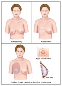
Mastectomy is surgery to remove the whole breast. (If you choose this option, you might have to decide whether to have surgery to reconstruct your breast and when.)
Breast-conserving surgery (also called “lumpectomy”) is surgery to remove the cancer and a section of healthy tissue around it. People who choose this option keep their breast. But they usually must have radiation therapy after surgery. - Radiation therapy – Radiation kills cancer cells.
- Chemotherapy – Chemotherapy is the medical term for medicines that kill cancer cells or stop them from growing. Some people take these medicines before surgery to shrink the cancer and make it easier to remove. Some take these medicines after surgery to keep cancer from growing, spreading, or coming back.
- Hormone therapy – Some forms of breast cancer grow in response to hormones. Your doctor might give you treatments to block hormones or to prevent your body from making certain kinds of hormones.
- Targeted therapy – Some medicines work only on cancers that have certain characteristics. Your doctor might test you to see if you have a kind of cancer that would respond to this kind of therapy.
- Immunotherapy – This is the term doctors use for medicines that work with the body’s infection-fighting system to stop cancer growth. Immunotherapy might be used with chemotherapy to treat certain types of advanced breast cancers.
Getting treated for breast cancer involves making many choices. Besides choosing which surgery to have, you might have to choose which medicines to take and when.
Always let your doctors and nurses know how you feel about a treatment. Any time you are offered a treatment, ask:
- What are the benefits of this treatment? Is it likely to help me live longer? Will it reduce or prevent symptoms?
- What are the downsides to this treatment?
- Are there alternatives to this treatment?
- What happens if I do not have this treatment?
A sentinel lymph node biopsy for breast cancer is an operation to check if your breast cancer has spread to your lymph nodes.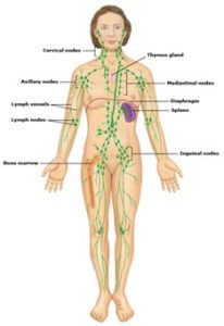
ImageLymph nodes are bean-shaped organs found all over the body, including the armpits, neck, and groin. They are part of a network of vessels called the called the “lymphatic system,” which carries a clear fluid called “lymph”. When breast cancer cells spread, they usually travel first through the lymph to 1 or more lymph nodes in the armpits. These lymph nodes are called “sentinel lymph nodes.”
During a sentinel lymph node biopsy a surgeon finds, takes out, and checks the sentinel lymph node (or nodes) for cancer cells. Knowing whether cancer is in the sentinel lymph nodes helps your doctor choose the right treatment for your breast cancer.
A sentinel lymph node biopsy is also called an “SLNB.”
The operation is usually done under general anesthesia. This means you will get medicines through a small tube into a vein (called an “IV”) or gases that you breathe through a mask to make you unconscious.
Doctors use 2 different techniques to find the sentinel node:
- Blue dye – For this technique, the doctor injects blue dye into your breast near the cancer after you are unconscious in the operating room. The dye travels through the lymph and turns the sentinel node blue.
- A radioactive substance – For this technique, the doctor injects a radioactive substance into your breast near the cancer and the colored area around your nipple, before you go to the operating room. Then, after you are unconscious, the doctor uses a special device that finds radioactive substances to locate the sentinel lymph node.
Most surgeons use both of these techniques, but some use just 1. After finding the sentinel node, the surgeon makes a small cut to take it out, then sends it to a lab. A different doctor then checks the node for cancer cells. Often, more than 1 sentinel node is found and taken out.
Most people do not have cancer in their sentinel lymph nodes. But if they do, the surgeon might take out other lymph nodes in the armpit to check them as well. If this happens, it is usually during a separate operation.
After an SLNB, you might have pain, bruising, bleeding, or get an infection. Your doctor will prescribe pain relievers or other medicines to treat any symptoms.
If your doctor used blue dye, part of your breast will be blue until all of the dye disappears from your body. Your urine will also turn green for 1 day.
In rare cases, people are allergic to the dye used for SLNB. Your doctor will tell you about this risk and answer any questions you have before the operation. The allergic reaction happens in the operating room and is treated right away with medicines given through an IV.
If your doctor used a radioactive substance, the amount of radioactivity is very low and not harmful. It leaves your body quickly through your urine. You are not “radioactive” or dangerous to other people near you.
Your doctor will let you know about the result of the biopsy. They will also tell you if additional lymph nodes need to be removed from your armpit.
- “Local treatment” – This is treatment directly to the breast with surgery or radiation.
- “Systemic treatment” – This is the name for medicines that treat the whole body. Chemotherapy is one example. There are other medicines that can be used, too. They destroy cancer cells or stop them from growing. They also lower the chances that the cancer will return anywhere in your body.
- Mastectomy – This is surgery to remove the whole breast. Most people who have a mastectomy can also have surgery to reconstruct the breast that is removed.
- Breast-conserving therapy (also called “lumpectomy”) – This is surgery to remove the cancer and a section of healthy tissue around it (called a “margin”). People who choose this option keep their breast. But they usually must have radiation therapy after surgery to kill any cancer cells that might still be in the breast area.
No. Studies show that people who choose lumpectomy live just as long as those who choose mastectomy.
| Mastectomy | Lumpectomy |
How will I look? | If you decide not to have your breast reconstructed, you will have a scar where your breast used to be. If you do have your breast reconstructed, you will have a new breast made of an implant or out of skin, muscle, and fat taken from other parts of your body. Reconstructed breasts do not have as much sensation as breasts that have had a lumpectomy. | Your breast will have a scar and might have a dent or dimple where the tissue was removed. The treated breast might be a little smaller than the other breast. Still, it will feel more like a real breast than a reconstructed breast and have more sensation. |
What will my recovery from surgery be like? | You will stay in the hospital for 1 to 2 days (or a little longer if you have reconstruction done, too). You will go home with drains that must be emptied twice a day and that stay in for about 2 weeks. Your recovery will take 4 to 6 weeks. During that time, you will need to rest and avoid sports, swimming, and heavy lifting. You might need physical therapy. | You will go home from the hospital the same day as your surgery, and your recovery will take 1 to 2 weeks. During that time, you will need to rest and avoid sports, swimming, and heavy lifting. You might need physical therapy. |
What are the risks or side effects of this surgery? | Mastectomy is usually safe, but it can sometimes lead to bleeding, infection, or fluid build-up. Plus, the surgery can sometimes damage the skin on the chest. (This is more common in people who smoke.) | Lumpectomy is usually safe, but it can sometimes lead to bleeding, infection, or fluid build-up where the cancer was removed. |
Will I need more than 1 surgery? | Possibly, if you decide to have your breast reconstructed later. | Possibly, if the cancer comes up to the margins and the surgeon has to take more tissue. |
Will I need radiation therapy? | Probably not. But some people who have mastectomy do need radiation. | Yes. After lumpectomy, most people must have radiation therapy 5 days a week for 3 to 6 weeks. |
What are the chances that cancer will come back in the same area? | Cancer can still recur after mastectomy because all the breast tissue cannot be totally removed. About 5 out of 100 people who have a mastectomy will have cancer come back on the chest wall after mastectomy. | Fewer than 10 out of 100 people who have a lumpectomy will have cancer recur. |
- Have more than 1 tumor in different areas of the same breast, which cannot be removed with a single cut
- Have cancer that has spread throughout the breast tissue, and the surgeon can’t get all the cancer out with a margin of normal tissue around it
- Cannot have radiation therapy, for example, because you are pregnant or have certain skin conditions
- Already had radiation to that area of your breast. (More radiation to the same area can cause too much damage.)
Coosing between mastectomy and lumpectomy
- The way you will look – Is it important to you to keep your own breast? How would you feel if your chest was flat or you had to wear a plastic breast in your bra? Ask to see pictures of people who have had the different kinds of surgery. Remember, if you choose mastectomy, can choose to have your breast reconstructed if you want. If you have small breasts and a large tumor, a lumpectomy might make your breast look quite different. In this case, mastectomy with reconstruction might be a better option. Some people do not mind having a flat chest after mastectomy, and choose not to have reconstruction.
- The risk that the cancer will come back – People who choose lumpectomy live just as long as those who choose mastectomy. They also have about the same chances of cancer returning in the breast. Most people whose cancer comes back after lumpectomy go on to have a mastectomy.
- The time involved and the side effects of radiation – After lumpectomy, most people must have radiation therapy. This usually involves getting treatments 5 days a week for 3 to 6 weeks, depending on the person’s age and other factors, although shorter treatments may be appropriate in some cases.
This type of radiation will not make your hair fall out, but it might make you tired. It will tan and possibly even burn the skin on your chest. This burn is like a sunburn and goes away fairly quickly. Toward the end of treatment, radiation can make you feel a little tired, but this is not very common and usually doesn’t last long. - Recovery from surgery
If you have a lumpectomy, you can go home from the hospital the same day as your surgery. For a week or 2 after surgery, you will need to rest and avoid sports, swimming, and heavy lifting.
If you have a mastectomy, you will stay in the hospital for 1 to 2 days after surgery. If you also have breast reconstruction, you might stay a day or 2 longer. You will go home with drains that must be emptied twice a day. After about 2 weeks, the surgeon will take the drains out in the office. If more fluid or blood builds up after the drains come out, the doctor will drain it with a needle in the office. During the draining process and for a few weeks more, you will need to rest and avoid sports, swimming, or heavy lifting. Even after you recover, you will not have normal feeling in your chest after a mastectomy. You can choose to have your breast reconstructed right away or later, or not at all.
No matter which surgery you have, you will probably need to have biopsies of your lymph nodes. This part of the surgery usually does not cause problems and is often done at the time of your breast surgery. But in some cases it can cause arm swelling, pain, or stiffness; shoulder pain or stiffness; or a nerve injury. If any of these happen to you, you might need to do special exercises or work with a physical therapist (exercise expert) to get back to normal.
People who have early-stage cancer or are having a mastectomy to prevent cancer can have the reconstruction at the same time as their mastectomy. This is called “immediate reconstruction.”
People with a later-stage or large cancer sometimes need to have radiation after mastectomy. (Radiation is a treatment that kills cancer cells or stops them from growing.) Reconstruction might be delayed until the radiation treatment is finished. This is called “delayed reconstruction.” The delay is needed because radiation could damage the reconstructed breast. There is also concern that an implant could keep radiation from reaching the right areas.
- How big your breasts are to begin with.
- If you are overweight and whether it affects your health – Your options will also depend on how much extra body fat you have and where it is.
- Whether you have other health problems – Some types of reconstruction are not recommended for people with diabetes, heart or lung disease, or other problems.
- Whether you have had surgery before – Scar tissue from surgery might affect your options, such as where donor tissue might be able to be taken from.
- Whether radiation therapy is planned – Some types of breast reconstruction are more likely to have problems related to radiation therapy.
- Your personal preferences – Some people prefer a shorter surgery and recovery time and choose implants for this reason. Some people do not wish to have man-made materials in their body and choose flaps.
A breast implant is basically a breast-shaped container that is filled with salt-water (called “saline”) or something that feels like Jell-O (called “silicone”). The implant can be inserted partly or completely under a layer of muscle in the chest.
Getting an implant usually involves 2 steps. First, the surgeon inserts a device called an “expander.” Often another piece of material (called “ADM”) is added to support the expander, and later the implant. The expander stretches the skin and muscle in the chest. The surgeon gradually adds more and more fluid to the expander until the skin and muscle are stretched enough for the size of the implant being used. Then, the surgeon does another surgery to replace the expander with the implant. Implants are best for people with smaller breasts that don’t droop.
A flap reconstruction uses tissue from another part of the body to create a new breast. That tissue might be rotated in place keeping its own blood supply (called a “pedicled” flap) or disconnected and then reattached to a new blood supply (called a “free” flap). How complicated the procedure is, and the risks involved, depend on the type of tissue used.
If you choose a flap, your options might include:
- Perforator flaps – A perforator flap is the most common type of flap used for breast reconstruction. It is usually taken from the belly. One type is called a “DIEP flap”. The flap is made up of skin and fat, but not muscle. After surgery, the belly looks flatter, like it does after a “tummy tuck.” Since there is no muscle removed, the belly is not weakened after this kind of flap.
For people who do not have enough belly fat, there might be other perforator flap options.
- TRAM flaps – A TRAM flap is also taken from the belly, but is made up of skin, fat, and muscle.Because the muscle is taken, the belly is weaker after surgery. There might also be a noticeable bulge where the muscle was removed, especially if both breasts are reconstructed with this type of flap.
- LD flap – A latissimus dorsi (LD) flap is taken from the back and is made up of skin, fat, and muscle. People who have this kind of flap have a scar on their back, just beneath the level of the bra line. They often also get an implant, because there is not enough fat on the back to make a new breast. An LD flap is more often used when other options are not available, or if a past reconstruction has not worked out.
If you want it to be, yes. This is usually done a few months after the breast reconstruction is done. To make a new nipple, the surgeon can rearrange the tissue that is already there or use tissue from another part of the body. Surgeons can also tattoo the new nipple and area around the nipple (called the areola) to match the color of your other nipple and make it look three-dimensional.
As much as possible, yes. But the new breast will never be exactly like the breast you had before or exactly like your other breast. Plus, you won’t have normal feeling (sensation) in the new breast. If you don’t like the difference between them, your surgeon might be able to make changes to the reconstructed breast, or operate on your other breast to make them look more similar.
Maybe. Only some of the reconstruction types will be appropriate for you. But if you think you would rather have 1 type of reconstruction over another, ask your surgeon if that approach would work for you. They can tell you if your choice makes sense, and if not, why not.
Most people do not have serious problems after breast reconstruction. But there are some problems that can happen, either right after the surgery or later on:
- If you had either type of reconstruction (implant or flap) – Problems can include infection, blood or fluid coming from the area where you had surgery, or pain that does not go away.
- If you had an implant– The most common problem is called “capsular contracture.” This is when scar tissue around the implant becomes hard and tight. This can cause the breast to feel firm or sore, or change shape. Other possible problems include the implant deflating, bursting, or moving out of place.
In very rare cases, people with an implant can get a certain type of lymphoma years later. Lymphoma is a cancer of the lymphatic system, which is part of the body’s immune (infection-fighting) system. If you have an implant, it’s important to see your doctor regularly afterwards. This way they can check for, and treat, any problems. - If you had a flap reconstruction– In some cases, the flap does not get enough blood. This can happen right after surgery, or sometimes later, and if it does, some or all of the flap might need to be removed. In this case, your surgeon will need to do more surgery to fix the reconstruction, or do another type or reconstruction. Some people can develop a bulge or hernia where the flap tissue was removed to make the flap.
Call your doctor or nurse if you think you have any of these problems. Some of them need treatment right away.
Abdomen
When you have a stomach ache, you have pain or discomfort in your belly. Sometimes that’s the only symptom you have. Other times, you can have other symptoms such as:
- Burning in your chest known as heartburn
- Burping
- Bloating (feeling like your belly is filled with air)
- Feeling full too quickly when you start eating
Most people do not need to see a doctor or nurse for a stomach ache. But you should see your doctor or nurse if:
- You have bloody bowel movements, diarrhea, or vomiting
- Your pain is severe and lasts more than an hour or comes and goes for more than 24 hours
- You cannot eat or drink for hours
- You have a fever higher than 102°F (39°C)
- You lose a lot of weight without trying to, or lose interest in food
In some cases, stomach aches are caused by a specific problem, such as:
- A stomach ulcer, which is a sore on the inside of the stomach
- A condition called “diverticulitis,” in which small pouches in your large intestine get infected
In other cases, doctors do not know what causes stomach aches or the other symptoms that happen with them. Even so, there are usually ways to treat the symptoms of stomach ache.
If your symptoms are caused by a specific problem, such as an ulcer, treating that problem will likely relieve your symptoms. But if your doctor or nurse does not know what is causing your pain, they might recommend medicines that reduce the amount of acid in your stomach. These medicines often relieve stomach ache and the symptoms that come with it. Some of these medicines are available without a prescription.
Yes. The foods you eat and the way you eat them can have a big effect on whether or not you feel pain.
To lower your chances of getting a stomach ache:
- Avoid fatty foods, such as red meat, butter, fried foods, and cheese
- Eat a bunch of small meals each day, rather than 2 or 3 big meals
- Stay away from foods that seem to make your symptoms worse
- Avoid taking over-the-counter medicines that seem to make your symptoms worse – Examples include aspirinor ibuprofen(sample brand names: Advil, Motrin).
Some people – especially kids – sometimes get a stomach ache after drinking milk or eating cheese, ice cream, or other foods that have milk in them. They have a problem called “lactose intolerance,” which means that they cannot fully break down foods that have milk in them.
People with lactose intolerance can avoid problems caused by milk if they take something called lactase. Lactase (sample brand name: Lactaid) helps your body break down milk. Some foods come with it already added.
If your stomach ache seems to be related to constipation, meaning that you do not have enough bowel movements, you might need more fiber or a medicine called a laxative. (Laxatives are medicines that increase the number of bowel movements you have.)
Taking in a lot of fiber helps to increase the number of bowel movements you have. You can get more fiber by:
- Eating plenty of fruits, vegetables, and whole grains
- Taking fiber pills, powders, or wafers
In general, yes. Children get stomach aches for most of the same reasons that adults do. But in children, stomach pain is often triggered by stress or anxiety. For them, it’s especially important to pay attention to psychological or emotional problems that might be making pain worse.
Yes. “Abdominal pain” means pain in the abdomen (or belly), which is the part of the body between the chest and the genital area. This pain can happen for different reasons. It can be “chronic,” which means it develops over time, or “acute,” which means it starts suddenly. It can be mild or severe. A person might feel the pain all over their abdomen, or only in 1 part.
Abdominal pain can feel different for different people. It can feel sharp or crampy, or dull and steady. Some people feel better if they curl into a ball, while others need to lie flat and completely still. People often feel sick to their stomach and retch or vomit.
Doctors use the term “acute abdomen” to describe an episode of severe abdominal pain that starts suddenly and lasts for a few hours or longer. It can cause pain so severe that the person has a hard time moving or breathing and it makes them want to go to the hospital or see their doctor or nurse right away. A true acute abdomen is a medical emergency.
Lots of different things can cause abdominal pain. When pain is less severe, it can be due to something like a virus or a stomach inflammation (called “gastritis”).
Acute pain that is more severe can be caused by problems with 1 or more organs in the abdomen. Organs in the abdomen can be part of the digestive, urinary, or reproductive systems.
Conditions that affect organs in the chest or genital area can also cause pain. Even though these organs aren’t in the abdomen, people might still have abdominal pain.
Common causes of acute abdominal pain in adults include:
- Appendicitis – Appendicitis is the term for when the appendix (a long, thin pouch that hangs down from the large intestine) gets infected and inflamed.
- Diverticulitis – Diverticulitis is an infection that develops in small pouches that can form in the intestine. This is common in older people.
- Gallstones – Gallstones are small stones that form inside an organ called the gallbladder, which stores bile, a fluid that helps the body break down fat.
- Abscess – An abscess is a collection of pus. An abscess can form in the abdomen, typically near the intestine.
- Kidney stones – Kidney stones can form when salts and minerals that are normally in the urine build up and harden. They can cause pain when they pass through the ureters, which are the tubes that carry urine from the kidney to the bladder.
- Bowel perforation – This is a hole in the bowel wall.
- Perforated ulcer – This is a hole in the wall of the stomach or intestine.
- Pancreatitis – This is the term for when the pancreas gets inflamed.
- Ruptured cyst in the ovary – Cysts in the ovary are fluid-filled sacs that can form in some women. They sometimes rupture, which means that they break open and spill out.
- Ectopic pregnancy – An ectopic pregnancy is a pregnancy that develops outside the uterus.
Yes. If you have sudden or severe abdominal pain, call your doctor or nurse or go to the hospital right away. Depending on the cause of your pain, you might need immediate treatment.
Probably. The doctor or nurse will ask about your symptoms, including where your pain is and what it feels like. The location of the pain can be an important clue to the cause.
Your doctor will ask about your current and past medical conditions, and do a physical exam. They might do repeat exams over time to follow your symptoms.
Your doctor will decide which tests you should have based on your symptoms and individual situation. The tests might include:
- Blood tests
- Urine tests
- X-rays
- An ultrasound, CT scan, or other imaging test – Imaging tests create pictures of the inside of the body.
Treatment depends on what’s causing the pain. It might include 1 or more of the following:
- Fluids given by IV
- Pain medicines
- Antibiotic medicines to treat an infection
- Other medicines to treat other medical conditions
- Surgery
Constipation is a common problem that makes it hard to have bowel movements. Your bowel movements might be:
- Too hard
- Too small
- Hard to get out
- Happening fewer than 3 times a week
Constipation can be caused by:
- Side effects of some medicines
- Poor diet
- Diseases of the digestive system
These symptoms could signal a more serious problem:
- Blood in the toilet or on the toilet paper after having a bowel movement
- Fever
- Weight loss
- Feeling weak
It could also be a sign of a problem if you have new constipation without a change in your medicines or diet, and have never had constipation in the past.
Yes. Try these steps:
- Eat foods that have a lot of fiber. Good choices are fruits, vegetables, prune juice, and cereal.
- Drink plenty of water and other fluids.
- When you feel the need to go to the bathroom, go to the bathroom. Don’t hold it.
- Take laxatives. These are medicines that help make bowel movements easier to get out. Some are pills that you swallow. Others go into the rectum. These are called “suppositories.”
See your doctor or nurse if:
- Your symptoms are new or not normal for you
- You do not have a bowel movement for a few days
- The problem comes and goes, but lasts for longer than 3 weeks
- You are in a lot of pain
- You have other symptoms that also worry you (for example, bleeding, weakness, weight loss, or fever)
- Other people in your family have had colorectal cancer or inflammatory bowel disease
Your doctor or nurse will decide which tests you should have based on your age, other symptoms, and individual situation. There are lots of tests, but you might not need any.
Here are the most common tests doctors use to find the cause of constipation:
- Rectal exam – Your doctor will look at the outside of your anus. They will also use a finger to feel inside the opening.
- Sigmoidoscopy or colonoscopy – For these tests, the doctor puts a thin tube into your anus. Then, they advance the tube into your large intestine. The large intestine is also called the colon. The tube has a camera attached to it, so the doctor can look inside your intestines. During these tests, the doctor can also take samples of tissue to look at under a microscope.
- X-rays, CT scan, or MRI – These create images of the inside of your body.
- Manometry studies – Manometry allows the doctor to measure the pressure inside the rectum at various points. It can help the doctor find out if the muscles that control bowel movements are working right. The test also shows whether the person’s rectum can feel normally.
That depends on what is causing your constipation. First, your doctor will want you to try eating more fiber and drinking more water. If that doesn’t help, your doctor might suggest:
- Medicines that you swallow or put in your rectum
- Changing the medicines you are taking for other conditions
- A treatment called an “enema” – For this treatment, a doctor or nurse will squirt water into your rectum. They might also use a thin tool to help break up bowel movements that are still inside you.
You might also be able to give yourself enema treatments at home, too. Enemas can be just water, or they can contain medicine to help with constipation. - Biofeedback – This is a technique that teaches you to relax your muscles so you can let go and push bowel movements out.
You can reduce your chances of getting constipation again by:
- Eating a diet that is full of fiber (table 1)
- Drinking water and other fluids during the day
- Going to the bathroom at regular times every day
Colon and rectal cancer screening is a way in which doctors check the colon and rectum for signs of cancer or growths (called polyps) that might become cancer. It is done in people who have no symptoms and no reason to think they have cancer. The goal is to find and remove polyps before they become cancer, or to find cancer early, before it grows, spreads, or causes problems.
The colon and rectum are the last part of the digestive tract. When doctors talk about colon and rectal cancer screening, they use the term “colorectal.” That is just a shorter way of saying “colon and rectal.” It’s also possible to say just colon cancer screening.
Studies show that having colon cancer screening lowers the chance of dying from colon cancer. There are several different types of screening test that can do this.
They include:
- Colonoscopy– Colonoscopy allows the doctor to see directly inside the entire colon. Before you can have a colonoscopy, you must clean out your colon. You do this at home by drinking a special liquid that causes watery diarrhea for several hours. On the day of the test, you get medicine to help you relax, if you want. Then a doctor puts a thin tube into your anus and advances it into your colon. The tube has a tiny camera attached to it, so the doctor can see inside your colon. The tube also has tiny tools on the end, so the doctor can remove pieces of tissue or polyps if they are there. After polyps or pieces of tissue are removed, they are sent to a lab to be checked for cancer.
- Advantages of this test – Colonoscopy finds most small polyps and almost all large polyps and cancers. If found, polyps can be removed right away. This test gives the most accurate results. If any other screening tests are done first and come back positive (abnormal), a colonoscopy will need to be done for follow-up. If you have a colonoscopy as your first test, you will probably not need a second follow-up test soon after.
- Drawbacks to this test – Colonoscopy has some risks. It can cause bleeding or tear the inside of the colon, but this only happens in 1 out of 1,000 people. Also, cleaning out the bowel beforehand can be unpleasant. Plus, people usually cannot work or drive for the rest of the day after the test, because of the relaxation medicine they take during the test.
In certain situations, a doctor might do something called a “capsule” colonoscopy. For this test, you swallow a special capsule that contains tiny wireless video cameras. This might be done if your doctor was not able to see all of your colon during a regular colonoscopy. - CT colonography(also known as virtual colonoscopy or CTC) – CTC looks for cancer and polyps using a special X-ray called a “CT scan.” For most CTC tests, the preparation is the same as it is for colonoscopy.
- Advantages of this test – CTC can find polyps and cancers in the whole colon without the need for medicines to relax.
- Drawbacks to this test – If doctors find polyps or cancer with CTC, they usually follow up with a colonoscopy. CTC sometimes finds areas that look abnormal but that turn out to be healthy. This means that CTC can lead to tests and procedures you did not need. Plus, CTC exposes you to radiation. In most cases, the preparation needed to clean the bowel is the same as the one needed for a colonoscopy. The test is expensive, and some insurance companies might not cover this test for screening.
- Stool test for blood– “Stool” is another word for bowel movements. Stool tests most commonly check for blood in samples of stool. Cancers and polyps can bleed, and if they bleed around the time you do the stool test, then blood will show up on the test. The test can find even small amounts of blood that you can’t see in your stool. Other less serious conditions can also cause small amounts of blood in the stool, and that will show up in this test. You will have to collect small samples from your bowel movements, which you will put in a special container you get from your doctor or nurse. Then you follow the instructions to mail the container out for the testing.
- Advantages of this test – This test does not involve cleaning out the colon or having any procedures.
- Drawbacks to this test – Stool tests are less likely to find polyps than other screening tests. These tests also often come up abnormal even in people who do not have cancer. If a stool test shows something abnormal, doctors usually follow up with a colonoscopy.
- Sigmoidoscopy– A sigmoidoscopy is similar in some ways to a colonoscopy. The difference is that this test looks only at the last part of the colon, and a colonoscopy looks at the whole colon. Before you have a sigmoidoscopy, you must clean out the lower part of your colon using an enema. This bowel cleaning is not as thorough or unpleasant as the one for colonoscopy. For this test, you do not need to take medicines to help you relax, so you can drive and work afterward if you want.
- Advantages of this test – Sigmoidoscopy can find polyps and cancers in the rectum and the last part of the colon. If polyps are found, they can be removed right away.
- Drawbacks to this test – In about 2 out of 10,000 people, sigmoidoscopy tears the inside of the colon. The test also can’t find polyps or cancers that are in the part of the colon the test does not view (figure 3). If doctors find polyps or cancer during a sigmoidoscopy, they usually follow up with a colonoscopy.
- Stool DNA test– The stool DNA test checks for genetic markers of cancer, as well as for signs of blood. For this test, you get a special kit in order to collect a whole bowel movement. Then you follow the instructions about how and where to ship it.
- Advantages of this test – This test does not involve cleaning out the colon or having any procedures. When cancer is not present, it is less likely to be falsely abnormal than a stool test for blood. That means it leads to fewer unnecessary colonoscopies.
- Drawbacks to this test – It might be unpleasant to collect and ship a whole bowel movement. If a DNA test shows something abnormal, doctors usually follow up with a colonoscopy.
There is no blood test that most experts think is accurate enough to use for screening.
Work with your doctor or nurse to decide which test is best for you. Some doctors might choose to combine screening tests, for example, sigmoidoscopy plus stool testing for blood. Being screened–no matter how–is more important than which test you choose.
Doctors recommend that most people begin having colon cancer screening at age 45. People who have an increased risk of getting colon cancer sometimes begin screening at a younger age. That might include people with a strong family history of colon cancer, and people with diseases of the colon called “Crohn’s disease” and “ulcerative colitis.”
Most people can stop being screened around the age of 75, or at the latest 85.
That depends on your risk of colon cancer and which test you have. People who have a high risk of colon cancer often need to be tested more often and should have a colonoscopy.
Most people are not at high risk, so they can choose one of these schedules:
- Colonoscopy every 10 years
- CT colonography (CTC) every 5 years
- Stool testing for blood once a year
- Sigmoidoscopy every 5 to 10 years
- Stool DNA testing every 3 years (but doctors are not yet sure of the best time frame for repeating the test)
A colonoscopy is a test that looks at the inner lining of a person’s large intestine. The large intestine is also called the colon.
Often, people have a colonoscopy as a screening test to check for polyps or for cancer in the colon or rectum. Polyps are growths in the colon that might turn into cancer. If you have polyps, the doctor can usually take them out during the colonoscopy. Taking polyps out lowers your chances of getting cancer. People can also have a colonoscopy if they have any of the symptoms listed below.
Cancer screening tests are tests that are done to try and find cancer early, before a person has symptoms. Cancer that is found early often is small and can be cured or treated easily.
Doctors can use 5 or 6 different tests to screen for colon cancer. But most doctors think that colonoscopy is the best test to screen for colon cancer.
Doctors recommend that most people begin having colon cancer screening at age 45. Some people have an increased chance of getting colon cancer, because of a strong family history or certain medical conditions. These people might begin screening at a younger age.
Your doctor might order a colonoscopy if you have:
- Blood in your bowel movements
- A change in your bowel habits
- A condition called anemia that can make you feel tired and weak
- Long-term belly or rectal pain that you cannot explain
- Abnormal results from a different type of colon test
- A history of colon cancer or polyps in your colon
Your doctor will give you instructions about what to do before a colonoscopy. They will tell you what foods you can and cannot eat. They will also tell you if you need to stop taking any of your usual medicines beforehand. Make sure to read the instructions as soon as you get them. You might have to stop some medicines up to a week before the test.
The colon needs to be cleaned out before a colonoscopy. Your doctor will give you a special drink that causes watery diarrhea. It is important to drink all of it to make sure your colon is clean. If your colon is clean, your doctor will get a better look at the inside lining of the colon. A clean colon also makes the test easier to do and more comfortable. Let your doctor know if you have trouble getting ready for your colonoscopy.
Your doctor will give you medicine to make you feel relaxed. Then they will put a thin tube with a camera and light on the end into your anus and up into the rectum and colon. Your doctor will look at the inside lining of the whole colon.
During the procedure, your doctor might do a test called a biopsy. During a biopsy, a doctor takes a small piece of tissue from the colon. Then they look at the tissue under a microscope to see if it has cancer. Your doctor might also remove growths that they see in the colon. You will not feel it if the doctor takes a biopsy or removes a growth.
Your doctor will give you instructions about what to do after a colonoscopy. Most people can eat as usual. But most doctors recommend that people do not drive or go to work for the rest of the day. Your doctor will tell you when to start taking any medicines you had to stop before the test.
Call your doctor or nurse immediately if you have any of the following problems after your colonoscopy:
- Belly pain that is much worse than gas pain or cramps
- A bloated and hard belly
- Vomiting
- Fever
- A lot of bleeding from your anus
Colon polyps are tiny growths that form on the inside of the large intestine (also known as the colon). Polyps are very common. About one-third to one-half of all adults have them by the time they are 50 years old. They do not usually cause symptoms. But some polyps can be or become cancer, so doctors sometimes remove them.
Colon polyps do not usually cause symptoms.
Doctors usually find colon polyps when they are doing screening tests to check for colon or rectal cancer. Cancer screening tests are tests that are done to try and find cancer early, before a person has symptoms. The screening tests for colon and rectal cancer include:
- Colonoscopy – Before having a colonoscopy, you will get medicine to help you relax. Then a doctor will put a thin tube into your anus and advance it into your colon. The tube has a camera attached to it, so the doctor can look inside your colon. The tube also has tools on the end, so the doctor can remove pieces of tissue, including polyps. After polyps are removed, they usually go to a lab to be tested for cancer and other problems.
- Sigmoidoscopy – A sigmoidoscopy is very similar to a colonoscopy. The only difference is that this test looks only at the first part of the colon, and a colonoscopy looks at the whole colon.
- CT colonography (also known as virtual colonoscopy) – For a virtual colonoscopy, you have a special kind of X-ray taken, called a “CT scan.” This test creates pictures of the colon.
- Stool test – “Stool” is another word for “bowel movements.” Stool tests check for blood or abnormal genes in samples of stool. If a stool test indicates that something might be wrong with the colon, doctors usually follow up with a colonoscopy. Then doctors find polyps, if they are there.
- Capsule colonoscopy – Rarely, your doctor might do something called a “capsule” colonoscopy. For this test, you swallow a special capsule that contains tiny wireless video cameras.
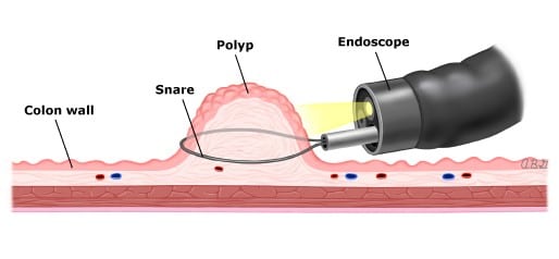 Doctors remove polyps using the same tools they use for a colonoscopy. They can remove polyps either by snipping them off with a special cutting tool, or by catching the polyps in a noose. Most polyps can be removed during a colonoscopy. But sometimes, large polyps need to be removed at a later time, either with another colonoscopy or with surgery.
You might need to have a colonoscopy every few years to check for more polyps. In some people polyps come back. And if you had the kind of polyps that could become cancer, your doctor will want to remove them as they appear. Also, if the polyps you had removed were the kind that could become cancer, people in your family might need to be checked for polyps and colon cancer earlier than if you did not have polyps.
Depending on your situation, your doctor might suggest genetic testing. This can show if your polyps are related to a specific gene that runs in families. If this turns out to be the case, they might recommend other tests that can be done to prevent cancer or find it early.
To reduce your chances of getting polyps or colon cancer:
- Eat a diet that is low in fat and high in fruits, vegetables, and fiber
- Lose weight, if you are overweight
- Do not smoke
- Limit the amount of alcohol you drink.
A colectomy is surgery in which your doctor removes part or all of your large intestine. The large intestine is also called the colon.
Doctors might do a colectomy to treat problems such as:
- Colon cancer
- Digestive tract disorders, such as severe diverticulitis or inflammatory bowel disease
- A blockage in the colon
- An injury to the colon
Your doctor will tell you how to prepare for your colectomy, and how long before the surgery you should stop eating and drinking.
Your doctor will tell you if you need to change or stop any of your medicines before the surgery. They might also give you medicine to take beforehand. For example, you will probably get antibiotics to prevent infection, plus a medicine to empty your intestines (this is called “bowel prep”).
Before the surgery starts, you will get medicines (through a thin tube that goes into a vein, called an “IV”) to make you sleep. You will not be awake for the surgery.
There are 2 main ways doctors can do a colectomy:
- Open surgery – During open surgery, your doctor will make a cut in your belly. They will remove some or all of your colon. How much your doctor removes depends on the reason for your surgery and how severe your condition is.
- Minimally invasive surgery – During minimally invasive surgery, your doctor will make a few small cuts in your belly. Then they will insert long, thin tools through the cuts and into your belly. One of the tools has a camera (called a “laparoscope”) on the end, which sends pictures to a TV screen. Your doctor can look at the screen to know where to cut and what to remove. Then they use the long tools to do the surgery through the small cuts. Sometimes they use a special robot to help move the tools.
After your doctor removes your colon, they will make sure there is a way for bowel movements to exit your body. To do this, your doctor will either:
- Reconnect your intestine – If your doctor can reconnect your intestine, you should be able to have bowel movements normally.
- Do a procedure called a “colostomy” or “ileostomy” – For either of these procedures, your doctor will make a small hole in your belly. Then they will connect your intestine to this opening. If your doctor connects your large intestine to the hole, it’s called a “colostomy.” If your doctor connects your small intestine to the hole, it’s called an “ileostomy.” Your bowel movements will come out through the hole into a bag that is attached to your skin.
An ileostomy is an opening in the belly made by a doctor as a way for waste products from the intestines to leave the body.
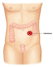 An ileostomy is sometimes called a “stoma,” which is a medical word that means “opening.” After you have an ileostomy made, waste products from your intestines will come out through the stoma into a bag that is attached to your skin.
A special nurse (called an ostomy nurse) will teach you how to take care of your ileostomy. They will teach you:
- When and how to empty the bag
- When and how to put on a new bag
- How to check your stoma for problems
There are different types of ileostomy bags that people can use. With some types of bags, you empty, clean, and reuse them. With other types, you throw them out after each use.
You might worry that your bag will leak, or that other people will be able to smell your bowel movements. But these things rarely happen. The bags are made so that they do not leak or smell.
Some people have a certain type of ileostomy. They don’t use a bag to collect their bowel movements. Instead, they have an internal pouch made out of intestine, which they drain through the stoma a few times a day.
Different problems can happen with an ileostomy, either right away or years later. Let your doctor or nurse know if you have any of the following symptoms or problems:
- Your stoma starts turning purple or black instead of pink.
- Your stoma is swollen or larger than usual or you have a bulge to the side of the stoma.
- Your stoma is smaller than usual.
- Your stoma leaks more than usual.
- You have a rash or sores around your stoma.
- You have diarrhea.
- You have sudden belly pain, cramps, or nausea.
- You are dehydrated. Dehydration is when the body loses too much water. Symptoms of dehydration include not making as much urine or having dark yellow urine, or feeling thirsty, tired, dizzy, or confused.
- You haven’t passed gas waste from your stoma for 4 to 6 hours (during the day). These symptoms could mean that your stoma is blocked.
When people have an ileostomy, their body doesn’t always absorb medicines normally. Because of this, try to use liquid medicines instead of pills. Do not take pills labeled “enteric coated,” “time release,” or “extended release.” Your body might not absorb these types of pills well.
Yes. When people have an ileostomy, their body doesn’t always absorb water, vitamins, and salts normally. Because of this, you should drink plenty of fluids to avoid getting dehydrated.
You should avoid eating foods that could easily block your intestine or stoma. Some of these foods include popcorn, mushrooms, dried fruit, and fruits or vegetables with skin.
The foods you eat can also affect the odor of your bowel movements, and how solid or loose they are. Certain foods can also make you have more gas. These are listed in the table.
Foods that can affect bowel movements and gas
|
|
|
|
You should be able to live an active and normal life with your ileostomy. But many people worry about the following things:
- Clothes – You do not need to wear special clothes. Other people won’t be able to see your bag under your clothes.
- Baths and showers – You can take a bath or shower with or without your bag on.
- Sports – You will probably be able to play most sports. You might want to wear a special belt to protect your bag and keep it in place. Doctors usually recommend that people with an ileostomy not play certain contact sports (such as football) or sports that involve straining, such as lifting weights.
- Swimming – You can swim with your bag on. Make sure to empty your bag before you swim.
- Sex – You can have sex. But you might want to wear a special wrap to protect (and cover) your bag during sex.
- Travel – When you travel, be sure to bring extra supplies for your ileostomy. If you fly, take your supplies in your carry-on luggage.
It is normal to feed sad, upset, or worried when you have an ileostomy. If you have these feelings, try to get help. You can talk with a family member, friend, or counselor. You might also find it helpful to go to a support group for people with an ileostomy.
A colostomy is an opening in the belly made by a doctor as a way for bowel movements to leave the body not through the anus. To make a colostomy, your doctor will do a procedure to make a small opening in your belly. Then they will connect your large intestine to this opening.
A colostomy is sometimes called a “stoma,” which is a medical word that means “opening.” After a colostomy is made, your bowel movements will come out through the stoma into a bag that is attached to your skin.
You will probably have about 4 to 8 bowel movements a day. Your bowel movements might be less solid than they used to be.
A special nurse (called an ostomy nurse) will teach you how to manage your colostomy. They will teach you:
- When and how to empty the bag that collects your bowel movements
- When and how to put on a new bag to collect your bowel movements
- How to check your stoma for problems
There are different types of colostomy bags. With some types of bags, you empty, clean, and reuse them. With other types, you throw them out after each use.
Some people worry that their bag will leak, or that other people will be able to smell their bowel movements. But this is not common. The bags are made so that they do not leak or smell.
If you have a certain type of colostomy, you might be able to manage it with a process called “irrigation.” This is a way to make your bowel movements regular. It involves squirting water into your stoma on a regular basis to
Food | Serving | Grams of fiber |
Fruits | ||
Apple (with skin) | 1 medium apple | 4.4 |
Banana | 1 medium banana | 3.1 |
Oranges | 1 orange | 3.1 |
Prunes | 1 cup, pitted | 12.4 |
Juices | ||
Apple, unsweetened, with added ascorbic acid | 1 cup | 0.5 |
Grapefruit, white, canned, sweetened | 1 cup | 0.2 |
Grape, unsweetened, with added ascorbic acid | 1 cup | 0.5 |
Orange | 1 cup | 0.7 |
Vegetables | ||
Cooked | ||
| 1 cup | 4.0 |
| 1/2 cup sliced | 2.3 |
| 1 cup | 8.8 |
| 1 medium potato | 3.8 |
Raw | ||
| 1 cucumber | 1.5 |
| 1 cup shredded | 0.5 |
| 1 medium tomato | 1.5 |
| 1 cup | 0.7 |
Legumes | ||
| 1 cup | 13.9 |
| 1 cup | 13.6 |
| 1 cup | 11.6 |
| 1 cup | 15.6 |
Breads, pastas, flours | ||
Bran muffins | 1 medium muffin | 5.2 |
Oatmeal, cooked | 1 cup | 4.0 |
White bread | 1 slice | 0.6 |
Whole-wheat bread | 1 slice | 1.9 |
Pasta and rice, cooked | ||
| 1 cup | 2.5 |
| 1 cup | 3.5 |
| 1 cup | 0.6 |
| 1 cup | 2.5 |
Nuts | ||
Almonds | 1/2 cup | 8.7 |
Peanuts | 1/2 cup | 7.9 |
Different problems can happen with a colostomy, either right away or years later. Let your doctor or nurse know if you have any of the following symptoms or problems:
- Your stoma is swollen or larger than usual.
- Your stoma is smaller than usual.
- Your stoma leaks more than usual.
- You have a rash or sores around your stoma.
- The inside of the stoma sticks out through the opening more than usual.
- You notice a bulge under the stoma or next to it.
- You have sudden belly pain, cramps, or nausea.
- You had a lot of diarrhea come out of the stoma and are dehydrated. Dehydration is when the body loses too much water. Symptoms of dehydration include not making as much urine or having dark yellow urine, or feeling thirsty, tired, dizzy, or confused.
- You haven’t had any gas or bowel movements through the stoma for 4 to 6 hours (during the day). These symptoms could mean that your stoma is blocked.
Probably not. But you should avoid getting constipated. (Constipation means trouble having bowel movements.) To avoid constipation, you can:
- Eat foods that have a lot of fiber
Amount of fiber in different foods
To learn how much fiber and other nutrients are in different foods, visit the United States Department of Agriculture (USDA) FoodData Central website.
- Drink plenty of water and other fluids
The foods you eat can affect the odor of your bowel movements, and how solid or soft they are. Certain foods can also make you have more gas. The foods that can affect your bowel movements and gas are listed in the table.
You should be able to live an active and normal life with your colostomy. But many people worry about the following things:
- Clothes – You do not need to wear special clothes. Other people won’t be able to see your bag under your clothes.
- Baths and showers – You can take a bath or shower with or without your bag on.
- Sports – You will probably be able to play most sports. You might want to wear a special belt to protect your bag and keep it in place. Doctors usually recommend that people with a colostomy not play certain contact sports (such as football) or lift weights.
- Swimming – You can swim with your bag on. Make sure to empty your bag before you swim.
- Sex – You can have sex. But you might want to wear a special wrap to protect (and cover) your bag during sex.
- Travel – When you travel, be sure to bring extra supplies to manage your colostomy. If you fly, take your supplies in your carry-on luggage.
It is normal to feel sad, upset, or worried when you have a colostomy. If you have these feelings, try to get help. You can talk with a family member, friend, or counselor. You might also find it helpful to go to a support group for people who have a colostomy.
Appendicitis is the name for when the appendix gets infected and inflamed. If that happens, it can swell and, in some cases, burst. That’s dangerous, because a burst appendix can cause infection in the belly.
The usual symptoms include:
- Severe pain in the lower part of the belly, on the right side. (For many people, the pain starts near the belly button and then moves to the lower right side.)
- Loss of appetite
- Nausea and vomiting
- Fever
Some people can have different symptoms, such as:
- Stomach upset
- Having a lot of gas
- Irregular bowel movements
- Diarrhea
- Feeling ill
If you have appendicitis, your doctor might be able to diagnose it just by doing an exam. They can learn a lot about your condition by pressing on your belly and talking with you about your symptoms.
If your doctor is not certain after the exam, they can do imaging tests to see what is causing your symptoms. Imaging tests create pictures of the inside of your body. They might include:
- A CT scan, which is a special kind of X-ray
- An ultrasound, which uses sound waves to look inside your belly
Yes. Call your doctor or nurse right away if you have the symptoms listed above. The risk of your appendix bursting is much higher after the first 24 hours of symptoms. If the appendix bursts, the surgery to treat it will be more complicated.
The main treatment for appendicitis is surgery to remove the appendix. This surgery can be done in 2 ways:
- Open surgery– During an open surgery, the doctor makes a cut near the appendix that is big enough to pull the appendix through.
- Laparoscopic surgery– During laparoscopic surgery, the doctor makes a few cuts that are much smaller than those used in open surgery. Then they insert long, thin tools into the belly. One of the tools has a camera (called a “laparoscope”) on the end, which sends pictures to a TV screen. The doctor can look at the image on the screen to know where to cut and what to remove. Then they use the long tools to do the surgery.
If your appendix has burst, your surgery will probably be more complicated than it would be if it had not burst. Your doctor will need to wash away the material that spills out when an appendix bursts. As a result, your cuts might be larger or you might spend more time in surgery.
Yes. But your options will depend on whether or not your appendix has burst.
If your appendix has not burst, it’s possible to treat appendicitis with just antibiotics. But, without surgery, there is a chance your appendicitis will come back again. So surgery is still the best treatment in most cases, but talk with your doctor if you wish to try to avoid surgery.
If your appendix has burst, but it has been a few days since this happened and you are feeling well, your doctor might decide not to do surgery right away. That’s because the body sometimes forms a pocket around the appendix to block off the infection. In this case, your doctor will probably give you antibiotics and watch you carefully until you are well. But you will still need surgery later to remove your appendix. That’s because at least 1 in 10 people with a burst appendix have a tumor in their appendix that can only be removed by surgery.
If you are pregnant and think you have signs of appendicitis, make sure you tell your doctors that you are pregnant. Doctors use ultrasound or a test called an “MRI” to check for appendicitis in people who are pregnant.
Diverticulitis is a disorder that can cause belly pain, fever, and problems with bowel movements.
The food we eat travels from the stomach through a long tube called the intestine. The last part of that tube is the colon. The colon sometimes has small pouches in its walls. These pouches are called “diverticula.” Many people who have these pouches have no symptoms. Diverticulitis happens when these pouches develop a small tear also known as a “microperforation,” which then become infected and cause symptoms.
The most common symptom of diverticulitis is pain, which is usually in the lower part of the belly. Other symptoms can include:
- Fever
- Constipation
- Diarrhea
- Nausea and vomiting
Yes. There are a few tests your doctor can do to find out if you have diverticulitis. But tests are not always needed. If you do have a test, you might have a:
- CT scan– A CT scan is a kind of imaging test. Imaging tests create pictures of the inside of your body.
- Abdominal ultrasound– This test uses sound waves to create pictures of your intestines.
Diverticulitis is typically treated with antibiotics. You might also need to go on a clear liquid diet for a short time. If you only have mild symptoms, this might be all the treatment you need.
But if you have severe symptoms, or if you get a fever, you might need to stay in the hospital. There, you can get fluids and antibiotics through a thin tube that goes into your vein, called an “IV.” That way you can stop eating and drinking until you get better.
If you have a serious infection, the doctor might put a tube into your belly to drain the infection. In very bad cases, people need surgery to remove the part of the colon that is affected.
A few months after your infection has been treated, your doctor might recommend that you have a procedure called a colonoscopy. During a colonoscopy, the doctor can look directly inside your colon to get an idea of the number of diverticula in your colon and to find out where they are. At the same time, they can check for signs of cancer.
If you have had diverticulitis, it’s a good idea to eat a lot of fiber. Good sources of fiber include fruits, oats, beans, peas, and green leafy vegetables. If you do not already eat fiber-rich foods, wait until after your symptoms get better to start.
You do not need to avoid seeds, nuts, popcorn, or other similar foods.
A peptic ulcer is a sore that can form on the lining of the stomach or duodenum. The duodenum is the first part of the small intestine.
Some people with peptic ulcers have no symptoms. Other people can have symptoms that include:
- Pain in the upper belly – Ulcers in the stomach often cause pain soon after a person eats. Ulcers in the duodenum often cause pain or burning when a person’s stomach is empty.
- Bloating, or feeling full after eating a small amount of food
- Not feeling hungry
- Nausea or vomiting
All of these symptoms can also be caused by other conditions. But if you have these symptoms, let your doctor or nurse know.
Sometimes, peptic ulcers can lead to serious problems. These include:
- Bleeding – This can cause smelly and black-colored bowel movements, vomiting blood, or more rarely, bright red bowel movements.
- A hole in the wall of the stomach or duodenum – This can cause sudden and severe belly pain.
- Obstruction – This is a blockage of the intestine. It can cause a feeling of fullness, bloating, indigestion, nausea, vomiting, belly pain shortly after eating, and weight loss.
Common causes of peptic ulcers include:
- An infection in the stomach or duodenum – This kind of infection is caused by a type of bacteria called “Helicobacter pylori” or “H. pylori.”
- Medicines called “nonsteroidal antiinflammatory drugs” (NSAIDs) – NSAIDs include pain-relieving medicines such as aspirin, ibuprofen(sample brand names: Advil, Motrin), and naproxen(sample brand names: Aleve, Naprosyn).
Yes. If you have symptoms of a peptic ulcer, your doctor might do:
- Tests to check for H. pyloriinfection – Doctors can check for H. pylori infection by doing:
- Breath tests – These tests measure substances in a person’s breath after they have been given a special liquid to drink
- Lab tests that check a sample of a bowel movement for the infection
- A procedure called an “upper endoscopy” – During an upper endoscopy, a doctor puts a thin tube with a camera on the end into a person’s mouth and down into the stomach and duodenum. Then they check the lining of the stomach and duodenum for ulcers.
Treatment depends on the cause, but most peptic ulcers are treated with medicines.
People with H. pylori infection are often treated with 3 or more medicines for 2 weeks to get rid of the infection. This treatment can include:
- Medicine to reduce the amount of acid that the stomach makes
- Different types of antibiotics
Some people need to take medicines that reduce the amount of acid for a longer amount of time. Some people take these medicines for the rest of their life.
It is important to follow all your doctors’ instructions about taking your medicines. Let your doctor or nurse know if you have any side effects from your medicines.
People who have serious problems from their peptic ulcers might also need to be treated with surgery.
After treatment, people often have follow-up tests. These can include:
- Tests to check that the H. pyloriinfection has gone away
- An upper endoscopy to check that the peptic ulcer has healed
To help a peptic ulcer heal and to prevent future peptic ulcers, you can:
- Not smoke
- Not take NSAIDs (if possible)
Pancreatitis is a condition that can cause severe belly pain.
The pancreas is an organ that makes hormones and juices that help break down food. Pancreatitis is the term for when this organ gets irritated or swollen.
Most people get over pancreatitis without any long-lasting effects. But a few people get very sick.
There are many causes of pancreatitis. But most cases are caused by gallstones or alcohol abuse:
- Gallstones – Gallstones are hard lumps that form inside an organ called the gallbladder. Both the pancreas and the gallbladder drain into a single tube. If that tube gets clogged by a gallstone, neither of the organs can drain. When that happens, the fluids from both organs get backed up. That can cause pain.
- Alcohol abuse – People who drink too much alcohol for too long sometimes get alcohol-related pancreatitis. People with this form of pancreatitis usually start to feel pain 1 to 3 days after drinking a lot of alcohol or after they suddenly stop drinking. They usually also have nausea and vomiting.
There are a few blood tests that can help your doctor or nurse figure out if you have pancreatitis. It’s also possible that your doctor will order a special kind of X-ray called a “CT scan” of your belly to check if belly pain is due to pancreatitis or other conditions.
Pancreatitis is usually treated in the hospital. There, your doctor or nurse can give you fluids and pain medicines to help you feel better. If you cannot eat, they can give you food through a tube.
Some people with pancreatitis get an infection, which can be treated with antibiotics. Other possible problems caused by pancreatitis are fluid buildup around the pancreas or organ failure. Fluid buildup around pancreas often goes away on its own but sometimes needs to be drained or treated with surgery. Organ failure is usually handled by a team of doctors in intensive care.
Another important part of treatment is to get rid of the cause of the pancreatitis. If your pancreatitis is caused by gallstones, your doctor might need to treat them, too. People with pancreatitis from alcohol use must learn to give up alcohol to keep from getting pancreatitis again.
Gallstones are small stones that form inside the gallbladder. They can be tiny specks or get as big as the whole gallbladder, which can be up to 6 inches long.
Normally, the gallbladder fills with bile in between meals. Then, when you eat fatty foods, the gallbladder empties the bile into the intestine. Sometimes, though, gallstones clog the gallbladder and keep it from draining. Other times, gallstones just irritate the gallbladder. If the gallstones are pushed out of the gallbladder, they can keep the liver or pancreas from draining.
In most cases, gallstones do not cause any symptoms. When they do cause symptoms, gallstones can cause:
- Belly pain – Often on the right side just under the rib cage or in the middle top portion of the belly
- Pain in the back or right shoulder
- Nausea and vomiting
If you know that you have gallstones but have no symptoms, you probably will not need treatment. But if you start having symptoms, you should get treated. The symptoms can come and go, but they often get worse over time.
Not usually. In some cases they can lead to serious problems, including:
- Jaundice, a condition that turns your skin and eyes yellow
- Infection of the gallbladder
- Tears in the gallbladder, which can lead to death
- Inflammation of the pancreas (the pancreas is an organ that makes hormones and juices involved in food breakdown)
Yes, doctors can find out if you have gallstones by doing an imaging test, such as an ultrasound. An ultrasound is a painless test that uses sound waves to create a picture of your gallbladder.
Even if tests show that you have gallstones, that does not mean they are causing symptoms. Your doctor might need to do other tests to make sure your stones and your symptoms are related.
People with gallstones generally have 3 treatment options. They can have:
- No treatment– This option is best for most people who have no symptoms. If they start having symptoms, they can think about treatment then.
- Surgery to remove the gallbladder and the stones– Gallbladder surgery is routine in the United States. But it involves using anesthesia, so it has some risks. The surgery does not affect digestion very much. But about half the people who have surgery have mild symptoms afterward, including watery bowel movements, gas, or bloating. These symptoms usually get better. People who have their gallbladder removed do not need to worry about gallstones coming back.
- Treatment to get rid of the stones but keep the gallbladder– People who choose this approach can take medicines to break up gallstones. In rare cases, doctors can use with a device to break up and remove stones. Medicines only work with some stones, and even then they take time – months to years. The stones can also come back after these treatments.
The right treatment for you will depend on:
- How large your stones are
- Whether you have symptoms, and how bad the symptoms are
- How you feel about the treatment options
Ask your doctor or nurse how each treatment might affect you. Then work with them to find the treatment that makes the most sense for you.
Yes. You can try to keep yourself at a healthy weight. People who are overweight are more likely to get gallstones.
If you plan to lose weight quickly – even if you have never had gallstones – ask your doctor or nurse what you can do to keep from getting gallstones. Losing weight quickly – for example, through weight loss surgery – can lead to gallstones. But your doctor or nurse can give you medicines to keep that from happening.
The gallbladder is a small, pear-shaped organ that is tucked under the liver. It stores bile, a fluid that is made in the liver and helps the body break down fat. When you eat a meal that has fat in it, the gallbladder empties the bile into a tube called the “bile duct.” The bile duct carries the bile into the small intestine to help with digestion.
Gallbladder removal is surgery to remove the gallbladder. This surgery is also called “cholecystectomy.”
There are 2 main ways to remove the gallbladder:
- Laparoscopic surgery– This means the surgeon uses a “laparoscope,” a long, thin tube that has a light and a tiny camera on the end to see inside the body. (The laparoscope is sometimes called a “scope” for short.) For this type of surgery, the surgeon makes a few small incisions. Then they insert the scope through 1 of the incisions and other special tools through the other incisions. Next the surgeon uses the scope and the tools to do the operation. Most gallbladder removals in the US are done using laparoscopic surgery. Sometimes, though, open surgery is necessary because the gallbladder and bile duct are too infected or scarred to do laparoscopic surgery safely.
- Open surgery– This means the surgeon makes an incision in your belly big enough to do the surgery directly.
The most common reason is to treat gallstones. Gallstones are small stones that form inside the gallbladder. These stones can block the ducts that bile flows through. The stones can cause inflammation, pain, and other symptoms.
This article is about gallbladder removal to treat gallstones. People can also have gallbladder removal to treat cancer of the gallbladder. But if cancer is the reason for the surgery, it usually involves removing more than just the gallbladder.
Before the surgery:
- Your doctor will order blood tests to check if your liver is working normally.
- Your doctor will order an imaging test called an ultrasound, which uses sound waves to create pictures of the inside of your body. This test will show if you have gallstones and if the bile duct is enlarged or blocked.
- If the bile duct is blocked by a stone, your doctor might order a procedure called “ERCP.” ERCP stands for “endoscopic retrograde cholangiopancreatography.” During this procedure, a doctor slides a tube called an “endoscope” down your throat. The endoscope has a tiny camera and a light on the end. The doctor advances the tube past your stomach and into your intestine to the spot where the bile duct empties into the intestine. Then the doctor injects a special dye that shows up on X-ray into the ducts that leads to the gallbladder, liver, and pancreas and takes an X-ray. That way they can see where the dye goes. In some cases, the doctor might also use the endoscope to remove some gallstones or widen the duct opening so that small stones can pass through.
- Your doctor might give you antibiotics through a thin tube that goes into a vein, called an “IV,” to reduce the risk of infection during and after surgery.
If you have the surgery to treat gallstones, the main benefit is that it will make your symptoms go away.
The risks of the surgery are low, but they can include:
- Damage to other bile ducts near the gallbladder
- Bile leaks
- Bleeding
- Damage to the bowels
- Infection
- Leaving gallstones “trapped” in the bile duct (which would need to be removed with ERCP after surgery)
In some cases, a person will continue to have belly pain even after the gallbladder is removed.
Recovery is a little different depending on whether you have laparoscopic or open surgery.
- If you have laparoscopic surgery, you will probably be able to leave the hospital the same day you have surgery. But there is some chance you will need to stay overnight. Even though the cuts on the belly are small, the operation inside was the same as if you had open surgery. Your doctor will want you to rest and avoid heavy lifting, sports, and swimming for at least a week.
- If you have open surgery, you will probably stay in the hospital for 1 to 2 days. While there, do your best to start walking as soon as possible. Also, do the breathing exercises that your nurse recommends. After you go home, you should be able to do most of your normal activities, but you should avoid heavy lifting, sports, and swimming for a few weeks.
If you are taking narcotic pain medicine during recovery, you might get constipated. Take a stool softener to prevent this problem.
If you develop any the following symptoms in the weeks after surgery, call your doctor:
- Fever or chills
- Redness or swelling around the cuts from your surgery
- Nausea or vomiting
- Cramping or more severe belly pain
- Bloating (feeling like your belly is full of gas)
- Yellow skin or eyes
- Urine that is very dark in color
The surgery does not affect digestion very much. But about half the people who have surgery have mild symptoms afterward, including loose bowel movements, gas, or bloating. These symptoms usually get better.
Minimally invasive surgery is a type of surgery that uses special tools designed to decrease the size of incisions and reduce how much the body’s tissues get damaged.
One kind of minimally invasive surgery involves the use of a “scope,” a viewing device that allows surgeons to look inside the body without opening it up all the way. Another type, called “endovascular surgery,” uses X-rays to see inside the body while the surgeon uses special devices that fit inside the blood vessels. This article is about the type of surgery that involves scopes.
There are several different types of scopes, but they all work in about the same way. They consist of a long, thin tube with a tiny camera and a light on the end. The camera sends pictures of the inside of the body to a TV screen. When doing this type of surgery, the surgeon makes a small incision just big enough for the scope to fit through. They also make 2 or more other incisions that slim tools can fit through. These tools include clamps, scissors, and stitching devices, which the surgeon can control from outside the body. While looking at the picture on the screen, the surgeon uses those tools to do the operation.
There are lots of different types . Their names are based on the body parts that are involved. The scopes used for the different types of surgery are named that way, too:
- Thoracoscopes are used in the chest for “thoracoscopic surgery.” (“Thorax” is Greek for chest.) This type of surgery can be used to remove pieces of lung or to do certain types of heart surgery.
- Laparoscopes are used in the belly for “laparoscopic surgery.” (“Lapara” is Greek for the space between the bottom of the rib cage and the hips.) This type of surgery can be used to remove the gallbladder, appendix, or uterus, or to do lots of other different procedures.
- Hysteroscopes are used in the uterus and vagina for “hysteroscopic surgery.” (“Hystera” is Greek for uterus.) This type of surgery can be used to remove abnormal growths in the uterus, or to do a number of different procedures on the uterus and vagina.
- Arthroscopes are used inside joints for “arthroscopic surgery.” (“Arthron” is Greek for joint.) This type of surgery can be used to repair or rebuild joints in the knee, shoulder, and hip.
Some minimally invasive surgeries involve a surgical “robot,” which is a machine that the surgeon controls. This is called “robot-assisted minimally invasive surgery” or simply “robotic surgery.” The tools used in robotic surgery allows for more controlled movements than regular tools.
In general – but not always – this type of surgery makes recovery easier. That’s because:
- It usually involves several small wounds, rather than one big one
- The organs don’t get moved around as much
Despite all of the differences with regular surgery, minimally invasive surgery is still surgery. People who have it do have some pain, they do often need stitches, and they can develop infections or other problems because of the surgery.
No. Many procedures can now be done through a minimally invasive approach. But it’s not entirely up to the person to choose what type of surgery they will have. Whether or not a person can have this type of surgery will depend on:
- Whether there is a surgeon available with enough experience doing the type of surgery the person needs
- Why the person needs surgery. (As an example, people who need surgery to remove very large cancers cannot always have minimally invasive procedures.)
- What other health problems the person might have (As an example, people who have serious heart or lung problems cannot tolerate minimally invasive surgery.)
Even when a person starts out having minimally invasive surgery, there’s no guarantee that the surgery will stay that way. Sometimes surgeons start out doing minimally invasive surgery and then switch to open surgery because they find something unexpected. This doesn’t mean the surgeon has done anything wrong. It is usually done to protect the person’s safety.
A small bowel obstruction (SBO) is a condition in which the small intestine gets blocked. “Small bowel” is another term for the small intestine. In an SBO, air, fluid, and food get stuck in the intestine. They can’t move through the small intestine the way they normally would.
The intestine can be partly or completely blocked in an SBO. A complete block can lead to serious problems. That’s because:
- The intestine can get swollen from the trapped air, fluid, and food. This swelling can make the intestine less able to absorb fluid, which can lead to dehydration and kidney failure.
- If the intestine wall tears, the fluid in the intestine can leak out. This can cause a belly infection.
- When the intestine is blocked, the blood vessels that bring oxygen to the intestine can get blocked, too. Without blood, parts of the intestine can die.
In some cases, the small intestine looks blocked on tests even when it isn’t. This could be from either intestinal “ileus” or “pseudo-obstruction.” This article is only about real obstruction.
The most common causes of an SBO are:
- Past surgery in the belly – After surgery, scar tissue can form in the belly and press on the intestine.
- Hernias – A hernia is an opening in the muscle or tissue that covers the muscle. Part of the intestine can slide through that opening and get trapped in a hernia. A hernia inside the belly can also cause SBO.
- Twisting of the intestines
●Tumor – Tumors (cancerous and noncancerous) can grow inside or outside the intestine and block it.
Symptoms depend on where your intestine is blocked and how blocked it is. The most common symptoms are:
- Nausea and vomiting
- Belly pain
- Belly swelling and bloating
- Not being able to have a bowel movement or pass gas
Yes. Your doctor will ask about your symptoms and do an exam. If your doctor thinks that you have an SBO, they will do imaging tests of your belly and blood tests.
The imaging tests can include an X-ray, CT scan, or a series of X-rays called a “GI series.” For the CT scan and GI series, you might drink a liquid called “contrast” beforehand. The contrast will show up on the CT scan or X-rays. It can help the doctor see what’s causing the blockage.
Treatment depends on the blockage and your symptoms.
If you have an SBO, you will be treated in the hospital. Your doctor will give you fluids and medicines. You will not be allowed to eat or drink. You will also get something called a “nasogastric tube.” This is a thin tube that goes in your nose, down your esophagus, and into your stomach. The tube can suck up the fluid and air in your stomach. This will make your stomach feel better and help keep you from vomiting.
Most people will not need any other treatment. That’s because, many times, an SBO can get better on its own. This is often the case for a partial SBO, or an SBO that has happened after earlier belly surgery.
Some people will need surgery. You are likely to need surgery if you have a belly infection, you have a complete blockage, or your SBO does not get better with fluids and the nasogastric tube in a few days. You also need surgery if the SBO is caused by a hernia, a tumor, or a twist in the intestine.
During surgery, your doctor will remove what is causing the blockage and, if needed, any unhealthy parts of your small intestine. If your doctor removes the unhealthy intestine, they will usually reconnect your intestines so you will not need an ostomy bag. (An ostomy bag is a bag that attaches to the skin and collects bowel movements. It is used if the intestines cannot be reconnected after a surgery.)
Intussusception is a condition that can cause severe belly pain. It happens when one part of the intestine slides into another part of the intestine. Intussusception can happen with either the small or large intestine.
When one part of the intestine slides into another, it causes a blockage. When the intestine is blocked, air, fluid, and food get stuck. They can’t move through the intestine the way they normally would. This causes symptoms.
Intussusception happens most often in babies and children, especially those younger than 3 years old. In some cases, intussusception is caused by another medical condition. But in most cases, doctors don’t know what caused the intussusception.
Symptoms usually start suddenly. They can include:
- Severe belly pain – The pain comes in waves. At first, episodes of pain happen about every 15 to 20 minutes, but the episodes get closer over time. The pain usually makes children cry and pull their knees up into their belly.
- Vomiting
- Bloody bowel movements
- Being very sleepy and hard to wake up
Some children with intussusception have only 1 or 2 of these symptoms. Other children with a small intussusception do not have any symptoms, although this is uncommon. Their doctor might find that they have an intussusception when the child has a test for another reason. Children who have no symptoms might not need treatment.
Yes. Call your child’s doctor or nurse right away if your child has symptoms of an intussusception.
Yes. The doctor or nurse will ask about your child’s symptoms and do an exam. They will do an imaging test of the belly, such as an X-ray or ultrasound. Imaging tests create pictures of the inside of the body.
The doctor will do a few things to treat your child’s intussusception.
First, they will probably give your child fluids through a thin tube that goes into a vein, called an “IV.”
Then, the doctor will fix the intussusception. There are different ways to do this. They include:
- A procedure called “nonoperative reduction” – This is also sometimes called an “air enema” or “contrast enema.” It is not surgery. For this procedure, the doctor will put a tube into your child’s rectum and push air or a special fluid into the tube. The air or fluid will go into the rectum and through the intestine. As the air or fluid reaches the intussusception, it makes the stuck intestine slide back out of the other part of the intestine. To see what they are doing, the doctor will do an ultrasound or X-ray during this procedure.
Some children will get a “nasogastric tube.” This is a tube that goes into the nose, down the esophagus, and into the stomach. The tube will suck up the fluid and air in the stomach. This will help your child feel better and keep them from vomiting.
- Surgery – If the intussusception could not be fixed with the nonoperative reduction, or if the intussusception has caused problems, your child will probably need surgery.
Ischemic bowel disease is a condition in which there is not enough blood flow to the intestines. This happens when veins or arteries in the intestines are blocked. It can happen in the large intestine or the small intestine. It can cause belly pain, nausea, diarrhea, and other symptoms.
The symptoms include:
- Belly pain – This can be mild or severe. It can:
- Start suddenly, or happen over several days or years.
- Start about 1 hour after eating, and last about 2 hours. Eating a big meal can make this pain even worse.
- Nausea, vomiting, or both
- Blood in bowel movements – This can be bloody diarrhea
- Weight loss, when not trying to lose weight
- Eating problems, such as:
- Food fear – Not wanting to eat because pain starts after eating
- Feeling full too quickly when eating
Yes. The doctor or nurse will ask about the symptoms and do an exam. They will also order one or more tests. These can include:
- X-ray, CT scan, or ultrasound of the belly – These imaging tests create pictures of the inside of the body. They can help show the cause of symptoms.
- Blood tests – These can show signs of not enough nutrition. Or they might show that a different condition is causing symptoms.
- Tests called “endoscopy,” “upper endoscopy,” “sigmoidoscopy,” or “colonoscopy” (figure 2) – For these tests, the doctor puts a thin tube down your throat and into your stomach, or up your rectum and into your colon. The tube has a camera attached so the doctor can look inside your body. The tube also has tools attached to it, which the doctor can use to take samples of tissue. These samples can go to the lab to be checked for problems.
- A test called an “angiogram” – For this test, the doctor injects a dye into the vessels that carry blood to the bowel. This dye can be seen with an imaging test such as a CT, MR, or fluoroscopy (a moving X-ray). The test can show if arteries around the bowel are blocked. If they are, this could be keeping part of the bowel from getting enough blood.
- Laparoscopy – This is a procedure that the doctor does in the operating room. The doctor makes a small cut near the belly button. Then they put a small device called a “laparoscope” inside. The doctor looks through the laparoscope to see if they can find the cause of symptoms.
Treatment for ischemic bowel disease depends on:
- What is blocking the blood vessels
- What type of vessel is blocked, an artery (which brings blood to the bowel) or a vein (which drains blood from the bowel)
- Whether the symptoms started suddenly or came on over a long time
Most people with ischemic bowel disease need treatment in the hospital. In the hospital, the doctor can:
- Give you fluids and nutrition through a small tube that goes into a vein, called an “IV”
- Put a thin tube called a “nasogastric tube” in your nose, down your esophagus, and into your stomach. If there is extra fluid and air in your stomach or intestines, the tube can suck it up. This will make you feel better and help keep you from vomiting.
Other treatments can include:
- Antibiotics to treat infection
- A procedure or surgery to open a blocked artery
- Anti-clotting medicines (sometimes called “blood thinners”) to treat clots in the vessels
- Surgery to take out part of the bowel if it is not healthy
These treatments can help the bowel work correctly again.
Splenectomy is surgery in which your spleen is removed. The spleen is an organ in the upper left part of the belly. It is part of the body’s infection-fighting system, or “immune system.” It makes blood cells that help you fight infection. The spleen also filters the blood. It removes things that could cause problems, including damaged blood cells and bacteria.
You might have a splenectomy to treat problems such as:
- Damage to the spleen – If your spleen gets injured, it can cause serious internal bleeding. This most often happens because a person is hit in the belly area, for example, in a car accident or while playing a contact sport. If the spleen gets badly injured, it might have to be removed.
- Certain disorders that affect blood cells – Examples include:
- Immune thrombocytopenia or “ITP,” a disorder in which a person’s own immune system destroys their platelets (a type of blood cell that helps blood clot)
- Autoimmune hemolytic anemia, a disorder in which a person’s own immune system destroys their red blood cells
- Beta thalassemia major (a condition some people are born with), in which the body does not make enough red blood cells
Usually, doctors try other treatments for these problems before they consider doing a splenectomy.
●A spleen that is much larger than usual – Doctors call this “splenomegaly.” Certain cancers or non-cancerous tumors or cysts can sometimes cause this. If it leads to symptoms, such as pain, you might need to have your spleen removed.
If possible, you will need to get vaccines before your surgery. The spleen helps protect the body from infections, so you will be more likely to get some kinds of serious infections after your spleen is removed. Vaccines help prevent this. They work by teaching your body how to fight the germs that cause infections. The vaccines you’ll need include those that protect against certain infections, called pneumococcus, meningococcus, and Haemophilus influenzae type b (or “Hib”). You should also get the usual vaccines that doctors recommend for everyone, such as the flu vaccine.
The vaccines you get will depend on which ones you’ve already had and when you got them. Ideally, you will get your vaccines at least 2 weeks before your surgery. This might not be possible if your spleen needs to be removed right away.
Your medical team will tell how long before the surgery you should stop eating and drinking. They will also tell you if you need to stop taking any of your medicines or start any new medicines.
Before the surgery starts, you will get medicines (through a thin tube that goes into a vein, called an “IV”) to make you sleep. You will not be awake for the surgery.
There are 2 different ways a splenectomy can be done:
- Open surgery – During open surgery, the doctor will make a cut, often in the middle of your belly, take out your spleen, and close the cut with special staples or stitches. If your spleen is very large or has been badly injured, you will most likely need open surgery.
- Laparoscopic surgery – During laparoscopic surgery, the doctor will make a few very small cuts in your belly and remove the spleen through one of these cuts. This is done by putting long, thin tools through the cuts. One of the tools (called a “laparoscope”) has a camera on the end, which sends pictures to a video screen. The doctor can look at the screen to see inside your body.
It depends on which type of surgery you had. If you had open surgery, you will need to stay in the hospital for a few days. If you had laparoscopic surgery, you might be able to go home the same day or the next day.
If you didn’t get all your vaccines before your surgery, you will get them afterwards. You might also get antibiotics to help your body prevent infections, since you no longer have a spleen to do this. You will get instructions about getting help quickly if you have a fever.
Most people can go back to their daily lives once they recover. But it is important to be aware of certain problems that can happen. These include:
- Infection – The spleen helps protect the body from infections. After your spleen has been removed, your body has a harder time fighting off certain infections. Also, without a spleen, even minor infections are more likely to turn into a serious problem called “sepsis.” Sepsis is a serious illness that happens when an infection travels through the whole body. If some serious infections are not treated right away, you can die.
Getting all the vaccines your doctor recommends helps prevent many infections. But it’s still possible to get an infection if you’ve had your vaccines. Your medical team will work with you to plan for what to do if you notice any signs of infection.
Because any infection could cause serious illness or even death, you will probably get antibiotics to keep at home. This way, you can start taking them at the first sign that you might have an infection. Signs of infection can include:
- Fever (temperature higher than 100.4°F or 38°C)
- Chills or shivering
- Sore throat
- Cough
- Earache
- Stuffy nose or sinus pain
- Headache
- Feeling sleepy or confused
- Nausea, vomiting, or diarrhea
- Feeling dizzy, or like you might pass out
- A fast heartbeat
- Small purple-red dots on the skin, or unexplained bruises
Your doctor will talk to you about what to do if you have any of these symptoms. If you have symptoms that could mean a serious infection, like fever or chills, you will need to go straight to the nearest emergency department. Do this even if you do not feel very sick. In the emergency department, they can check you for infection and decide if you need more or different treatment. For less serious symptoms, your doctor might tell you to call their office for advice on what to do next.
Depending on your age and the reason your spleen was removed, your doctor might also tell you to take an antibiotic every day. This will help prevent infection.
- Blood clots – The risk of blood clots also goes up after splenectomy. If a blood clot happens, it is most often in the legs (called a “deep vein thrombosis” or “DVT”) or the lungs (called a “pulmonary embolism” or “PE”). Symptoms of DVT can include swelling, pain, or warmth and redness in the leg. Symptoms of PE can include trouble breathing, chest pain when you breathe in, or coughing.
There are things you can do to help lower your risk of blood clots. On a long car trip or airplane flight, get up and walk around or move your legs frequently, every hour or so if possible. If you have surgery, make sure to let the doctors know that your spleen was removed so they know about this risk.
Some hormones can also increase the risk of blood clots. If you plan to take a hormonal treatment, such as birth control pills, let the doctor or nurse prescribing it know that you have had a splenectomy.
It is important to carry an alert card, or wear a medical bracelet, so others know you do not have a spleen. This will help doctors and nurses give you the best care if there is ever an emergency.
Hemorrhoids do not always cause symptoms. But when they do, symptoms can include:
- Itching of the skin around the anus
- Bleeding – Bleeding is usually painless. You might see bright red blood after using the toilet.
- Pain – If a blood clot forms inside a hemorrhoid, this can cause pain. It can also cause a lump that you might be able to feel.
- Swelling – Hemorrhoids can swell or dangle outside of the rectum during a bowel movement.
You should see a doctor or nurse if you have any symptoms, especially bleeding or if your bowel movements look like tar. Bleeding could be caused by something other than hemorrhoids, so you should have it checked out.
If you do have hemorrhoids, your doctor or nurse can suggest treatments. But there some steps you can try on your own first.
The most important thing you can do is to keep from getting constipated. You should have a bowel movement at least a few times a week. When you have a bowel movement, you also should not have to push too much. Plus, your bowel movements should not be too hard, and you should not spend too much time on the toilet (for example, reading).
Being constipated and having hard bowel movements can make hemorrhoids worse. Here are some steps you can take to avoid getting constipated or having hard stools:
- Eat lots of fruits, vegetables, and other foods with fiber. Fiber helps to increase bowel movements.
You need 20 to 35 grams of fiber a day to keep your bowel movements regular. If you do not get enough fiber from your diet, you can take fiber supplements. These come in the form of powders, wafers, or pills. They include psyllium seed (sample brand names: Metamucil, Konsyl), methylcellulose (sample brand name: Citrucel), polycarbophil (sample brand name: FiberCon), and wheat dextrin (sample brand name: Benefiber). If you take a fiber supplement, be sure to read the label so you know how much to take. If you’re not sure, ask your doctor nurse.
- Take medicines called “stool softeners” such as docusatesodium (sample brand names: Colace, Dulcolax). These medicines increase the number of bowel movements you have. They are safe to take and they can prevent problems later.
Some people feel better if they soak their buttocks in 2 or 3 inches of warm water. You can do this up to 2 to 3 times a day for 10 to 15 minutes. Do not add soap, bubble bath, or anything to the water.
There are also medicines that you can get without a prescription. They are usually creams or ointments that you rub on your anus to relieve pain, itching, and swelling. Some hemorrhoid medicines come in a capsule (called a suppository) that you put inside your rectum. Others come in a cream that comes in a bottle with a nozzle that you put inside your rectum. It is OK to try these medicines. But do not use medicines that have hydrocortisone (a steroid medicine) for more than a week, unless your doctor or nurse approves.
If you still have symptoms after trying the steps listed above, you might need treatments to destroy or remove the hemorrhoids.
One popular treatment for hemorrhoids inside the rectum is called “rubber band ligation.” For this treatment, the doctor ties tiny rubber bands around the hemorrhoids. A few days later the hemorrhoids shrink and stop bleeding. Doctors can also use lasers, heat, or chemicals to destroy hemorrhoids. But if none of these options works, your doctor might suggest surgery to remove or tie off the blood vessels of the hemorrhoids. Hemorrhoids on the outside of the rectum can only be removed with surgery.
An anal fissure is a tear in the lining of the anus, the opening where your bowel movements come out. Anal fissures cause pain, especially during a bowel movement.
There is a muscle that wraps around the anus and holds it shut. It is called the “anal sphincter.” The sphincter gets tense when the anus is injured. In people with anal fissures, the sphincter goes into spasms, which can lead to further injury.
An anal fissure is most often caused by having a hard, dry bowel movement.
Most people who have an anal fissure feel a tearing, ripping, or burning pain when they have a bowel movement. This pain can last for hours. Some people also bleed slightly when they have a bowel movement. They might see bright red blood on the toilet paper or on the surface of the bowel movement. Some people with an anal fissure also have itching or irritation around the anus.
Yes. See your doctor or nurse if you bleed when you have a bowel movement.
Your doctor or nurse can check whether you have anal fissure by gently spreading your buttocks apart and looking at your anus.
If you have had bleeding, your doctor or nurse might send you for more testing after the fissure has healed. This involves a “sigmoidoscopy” or “colonoscopy” . For these tests, the doctor puts a thin tube into your anus and up into your colon. The tube has a camera attached to it, so the doctor can look inside your colon and check for causes of bleeding.
For the first month of treatment, doctors recommend trying to keep your bowel movements soft so they are easier to pass. To do this, you can:
- Take fiber supplements along with plenty of water. Supplements include:
- Psyllium(sample brand name: Metamucil)
- Methylcellulose(sample brand name: Citrucel)
- Calcium polycarbophil(sample brand name: FiberCon)
- Wheat dextrin(sample brand name: Benefiber)
- Take a stool softener if the fiber supplements are not enough. An example of a stool softener is docusate(sample brand name: Colace).
Your doctor can also prescribe a medicine that relaxes the anal sphincter muscle. This helps the fissure heal. The most commonly used medicine is nitroglycerin or nifedipine cream. You will need smear the cream around the fissure twice a day every day. It’s important to keep doing this for as long as your doctor tells you to, even if you do not have pain every day. This will allow your fissure to heal completely.
You can also soak your buttocks in 2 or 3 inches of warm water. This is called taking a “sitz bath.” Sitz baths can help relieve pain by relaxing the sphincter. If you find that Sitz bath helps, do it 2 to 3 times a day for 10 to 15 minutes. Do not add soap, bubble bath, or anything else to the water.
If these steps to do not work in 1 to 2 months, doctors can try other treatments, such as:
- Botulinum toxin (“BoTox”) – This is a shot that can help the anal sphincter muscle relax and heal. It can help, but it can also cause short-term problems with leaking of gas or bowel movements.
- Surgery – During surgery, the doctor makes a small cut in the sphincter to help it relax. This surgery works in most patients, but doctors offer it only to people who do not get better with other treatments. Surgery can cause lasting problems with leaking of gas or bowel movements.
ID and Other Questions
A provider that has had additional training to treat complicated conditions. For Physicians this is a 2 – 3 year period after finishing Internal Medicine training. They commonly deal with fungal infections, parasitic, Specific viral conditions and complicated bacterial infections.
Stewardship is one of the core values of infectious disease providers. They work to ensure the most appropriate antibiotic for the correct amount of time. Unlike other medications like blood pressure medication or pain medication, Antibiotic overuse hurts patients, hospital and communities. All of our ID providers are involved in some capacity with the hospital stewardship where they work.
Infectious disease providers are able to give specific recommendations on vaccines and prophylactic measures depending on what part of the world you’re going and what activities you will be performing. IF you’d like to have a travel evaluation feel free to make an appointment with our office staff.
The virus that causes Autoimmune Deficiency Syndrome (AIDS), our providers are trained or Certified by AAHIVM (American Association of HIV Medicine). Patients that have HIV properly managed have a lifespan and quality of life the same as people without HIV diagnosis. Feel free to contact one of our providers for an appointment.
Staph Aureus is a bacterium that normally lives in the skin of most people. At times these bacteria may find a portal of entry and cause chaos in the body of their host. Depending on the availability of antibiotics to treat this organism it can be divided in Methicillin resistant Staph Aureus (less options) and Methicillin Susceptible Staph Aureus (more options)
It’s an infection in the inner lining of the heart which commonly involves the heart valves. Without treatment, infectious endocarditis has a 100% mortality rate. On average, patients need to be diagnosed in the hospital and followed by a specialist post hospitalization.
It is an unusual infection caused by a very slow growing bacteria. It is unclear why some people have the disease, but it can be treated with a cocktail of antibiotics. Evaluation with an ID provider should be the first step since not all diagnosed cases may need to be treated. On average, treatment is more than 12 months
https://www.lung.org/lung-health-diseases/lung-disease-lookup/mac-lung-disease
Osteo-(bone)-myelitis is deep seated infection in the bone that commonly requires 4 weeks or more of treatment. Success rates for Osteomyelitis are dependent on patient factors, and bacterial factors. In our area foot ulcers are probably the most common route of osteomyelitis.
Disease cause by SARS-CoV-2 virus that has affected the world which is why it’s called a pandemic. Disinformation about this virus is rampant. In Regional Clinic we follow evidence-based medicine and recommend our patients to be vaccinated + booster
Some people develop a chronic condition of this virus called “long covid” or post covid syndrome. Research is still ongoing to understand this disease presentation and we are still actively looking for potential effective treatment.
This disease is spread through tick bites by a spirochete that may affect multiple systems (Central Nervous, Muscle, Joints, Heart). After the initial infection, some patients may persist having symptoms. Multiple studies have attempted to answer if additional antibiotics help in this condition. The best level of evidence indicates that antibiotics have no role to play after the initial infection.
Providers that offer prolonged antibiotic cocktails for this condition are doing it based on their personal experience. The Infectious Disease Society of America (IDSA) and American College of Physicians (ACP) do not encourage/vouch these types of treatments.
https://www.niaid.nih.gov/diseases-conditions/chronic-lyme-disease


